AVDC® Nomenclature
Determining and adopting nomenclature is an ongoing process. Additional items will be added to this page as they are approved by the Nomenclature Committee and AVDC® and Foundation for Veterinary Dentistry Boards. This webpage is not meant to be a veterinary dental textbook. It provides definitions for structures, diseases and treatment procedures relevant to the oral cavity. Its primary purpose to is provide definitive terms for use by AVDC® residents and diplomates to permit optimal communication in case logs and articles.
Abbreviations to be used in AVDC® Case Logs are shown in (brackets)
- General Definitions
- Anatomy of Oral, Dental and Related Structures
- Teeth Abnormalities and Related Procedures
- Jaw and TMJ Abnormalities
- Periodontal Anatomy and Disease
- Oral Pathology: Inflammatory Diseases, Tumors, Other Abnormalities
- Tongue, Lips, Palate, Pharynx, Nose, Face, Salivary Glands and Lymph Nodes
- Occlusal Abnormalities
- Oral Surgery
- Equine Specific Terminology
Definitions of Veterinary Dentistry, Equine Dentistry, and Beakology
Definitions of Items Applying to More than One Oral Tissue or Disease
——–
Definitions of Veterinary Dentistry, Equine Dentistry and Beakology
_________________________________________
Veterinary dentistry
Veterinary Dentistry is a discipline within the scope of veterinary practice that involves the professional consultation, evaluation, diagnosis, prevention, treatment (nonsurgical, surgical or related procedures) of conditions, diseases, and disorders of the oral cavity and maxillofacial area and their adjacent and associated structures; it is provided by a licensed veterinarian, within the scope of his/her education, training and experience, in accordance with the ethics of the profession and applicable law.
Equine dentistry
Equine Dentistry is the practice of veterinary dentistry performed in equids (genus Equus: horses, asses and zebras).
Beakology
Beakology is the branch of science dealing with the anatomy, physiology and pathology
(including diagnosis and treatment of such pathology) of the beak and associated tissues of
vertebrate animals that have beaks or beaklike structures.
Definitions of Items Applying to More than One Oral Tissue or Disease
_________________________________________
Congenital
Of or relating to a disease, condition or characteristic that is present at birth and may be inherited or result from an insult during pregnancy.
Acquired
Of or relating to a disease, condition or characteristic that develops after birth and is not inherited.
Inherited
Of or relating to a disease, condition or characteristic that results from the genetic makeup of the individual animal and may be present at birth or develop later in life.
Culture/sensitivity (CS)
Bacteria cultured in medium and analyzed for sensitivity to antibiotics.
Laceration (LAC)
A tear or cut in the gingiva/alveolar mucosa (LAC/G), tongue/sublingual mucosa (LAC/T), lip skin/labial mucosa (LAC/L), cheek skin/buccal mucosa (LAC/B), palatal mucosa (LAC/P), or palatine tonsil/oropharyngeal mucosa (LAC/O); debridement and suturing of such.
Chewing lesion (CL)
Mucosal lesion resulting from selfinduced bite trauma on the cheek (CL/B), lip (CL/L), palate (CL/P) or tongue/sublingual region (CL/T).
Foreign body (FB)
An object originating outside the body; removal of the foreign body is abbreviated with FB/R.
Burn (TMA/BRN)
Injury to skin, mucosa or other body parts due to fire, heat, radiation, electricity, or a caustic
agent
Ballistic trauma (TMA/B)
Physical trauma sustained from a projectile that was launched through space, most commonly by a weapon such as a gun or a bow.
Electric injury (TMA/E)
Physical trauma to skin, mucosa or other tissues when coming into direct contact with an electrical current.
Anatomy of Teeth
Numbering Teeth
Generations of Teeth
Surfaces and Directions in the Mouth
——–
Dental Anatomy
_________________________________________
Pulp cavity
Space within the tooth
Pulp chamber
Space within the crown of a tooth
Root canal
Space within the root of a tooth
Apical foramen
Opening at the apex of a tooth, through which neurovascular structures pass to and from the dental pulp
Apical delta
Multiple apical foramina forming a branching pattern at the apex of a tooth reminiscent of a river delta when sectioned and viewed through a microscope that occurs in some brachyodont teeth
Ameloblasts
Epithelial cells involved in the formation of enamel (amelogenesis)
Enamel (E)
Mineralized tissue covering the crown of brachyodont teeth
Anatomical crown (CR/AC)
That part of a tooth that is coronal to the cementoenamel junction (or anatomical root)
Clinical crown (CR/CC)
That part of a tooth that is coronal to the gingival margin; also called erupted crown in equines
Anatomical root (RO/AR)
That part of a tooth that is apical to the cementoenamel junction (or anatomical crown)
Clinical root (RO/CR)
That part of a brachyodont tooth that is apical to the gingival margin
Cementoenamel junction
Area of a tooth where cementum and enamel meet
Reserve crown (CR/RC)
That part of the crown of a hypsodont tooth that is apical to the gingival margin
Nomenclature and Numbering of Teeth
_________________________________________
Incisor Teeth
The incisors will be referred to as: (right or left) (maxillary or mandibular) first, second, or third incisors numbered from the midline. Reference: Peyer B. Comparative odontology. 1st ed. Chicago: University of Chicago Press, 1968;1-347. Nickel R, Schummer A, Seiferle E, et al. Teeth, general and comparative. In: The viscera of domestic mammals. 1st ed. Berlin: Verlag Paul Parey, 1973;75-99.
Premolar Teeth in the Cat:
In the cat, the tooth immediately distal to the maxillary canine is the second premolar, the tooth immediately distal to the mandibular canine is the third premolar.
Reference(s): Nickel R, Schummer A, Seiferle E, et al. Teeth, general and comparative. In: The viscera of domestic mammals. 1st ed. Berlin: Verlag Paul Parey, 1973;75-99.
Tooth Numbering:
The existence of the conventional anatomical names of teeth as well as the various tooth numbering systems is recognized. The correct anatomical names of teeth are (right or left), (maxillary or mandibular), (first, second, third or fourth), (incisor, canine, premolar, molar), as applicable, written out in full or abbreviated. The modified Triadan system is presently considered to be the tooth numbering system of choice in veterinary dentistry; gaps are left in the numbering sequence where there are missing teeth (for example, the first premolar encountered in the feline left maxilla is numbered 206, not 205. The two lower right premolars are 407 and 408, not 405 and 406).
Both the use of anatomical names and the modified Triadan system are acceptable for recording and storing veterinary dental information. The use of anatomical names in publications is required by many leading journals and is recommended. It offers the advantage of veterinary dental publications being understandable to other health professionals and scientists with an interest in veterinary dentistry.
Reference(s): Floyd MR. The modified Triadan system: nomenclature for veterinary dentistry. J Vet Dent 1991; 8:18-19.
Comments:
In January 1972, the International Dental Federation adopted a new, two digit, user friendly nomenclature system for use in the human dental patient. This new system eliminated the plus and minus signs of the Haderup System and the brackets of the Winkel System. Following the acceptance of the new system for human dental nomenclature, Professor DrMedDent H. Triadan, a dentist at the University of Bern, Switzerland, introduced a similar system for animals. Due to the fact that many animals, including his canine model, have more than nine teeth in a quadrant, the Triadan system for animals utilizes three digits instead of two digits.
Abbreviations associated with Teeth:
Tooth (T): Hard structure embedded in the jaw; used for biting and chewing
Incisor (I): Incisor tooth
Canine (C): Canine tooth
Pemolar (PM): Premolar tooth
Molar (M): Molar tooth
Alveolus (A): Socket in the jaw for a tooth root or reserve crown (plural: alveoli)
Crown (C): Coronal portion of a tooth
Root (RO): Radicular portion of a tooth
Apex (AP): End of the root or reserve crown (plural: apices)
Generations of Teeth in Diphyodont Species
_________________________________________
Deciduous and Permanent are the anatomically correct terms to denote the two generations of teeth in diphyodont species.
It is acceptable to use “primary“ instead of deciduous in communicating with clients.
Reference: Anonymous. Nomina Anatomica Veterinaria. 4th ed. Zurich and Ithaca: World Association of Veterinary Anatomists, 1994. Boucher CO, Zwemer TJ. Boucher’s clinical dental terminology – a glossary of accepted terms in all disciplines of dentistry. 4th ed. St. Louis: Mosby, 1993. Evans HE. Miller’s anatomy of the dog. 3rd ed. Philadelphia: WB Saunders Co, 1993.
Comments: Deciduous is the scientific term used in biology, as well as in comparative anatomy and anthropology for both animal and plant structures which are regularly shed. As a substitute for temporary, the term primary appeared early in the literature and it is listed in both Anthony’s and Otofy’s dictionaries 1922-23. The style of the Journal of the ADA requires the term deciduous in all literature designed for the profession and allows primary only in discourse for non-professional persons.
Deciduous tooth (DT):
Primary tooth replaced by a permanent (secondary) tooth.
The deciduous dentition period is that period during which only deciduous teeth are present.
The mixed dentition period is that period during which both deciduous and permanent teeth are present.
The permanent dentition period is that period during which only permanent teeth are present.
Reference: Anonymous. Nomina anatomica veterinaria. 4th ed. Zurich and Ithaca: World Association of Veterinary Anatomists, 1994. Boucher CO, Zwemer TJ. Boucher’s clinical dental terminology – a glossary of accepted terms in all disciplines of dentistry. 4th ed. St. Louis: Mosby, 1993. Evans HE. Miller’s anatomy of the dog. 3rd ed. Philadelphia: WB Saunders Co, 1993.
The term “Persistent deciduous tooth” is etymologically correct, although the term “retained deciduous tooth” is commonly used. The latter term, however, can be confused with an unerupted deciduous tooth.
Reference: Eisenmenger E, Zetner K. Tierv§rztliche Zahnheilkunde. 1st ed. Berlin: Verlag Paul Parey, 1982;44-50.
Surfaces of Teeth and Directions in the Mouth
_________________________________________
Vestibular/Buccal/Labial
Vestibular is the correct term referring to the surface of the tooth facing the vestibule or lips; buccal and labial are acceptable alternatives.
Reference(s): Anonymous. Nomina Anatomica Veterinaria. 4th ed. Zurich and Ithaca: World Association of Veterinary Anatomists, 1994.
Comment(s): The term “facial” specifically refers to the surfaces of the rostral teeth visible from the front. According to Dr. A.J. Bezuidenhout, a veterinary anatomist at Cornell University, “facial” is a bit of a misnomer. Traditionally “facial” has been used in human dentistry for the aspect of teeth visible from the front, i.e. incisors and canines.
Lingual/Palatal
Lingual: The surface of a mandibular or maxillary tooth facing the tongue is the lingual surface. Palatal can also be used when referring to the lingual surface of maxillary teeth.
Mesial/Distal
Mesial and distal are terms applicable to tooth surfaces. The mesial surface of the first incisor is next to the median plane; on other teeth it is directed toward the first incisor. The distal surface is opposite from the mesial surface.
Rostral/Caudal
Rostral and caudal are the positional and directional anatomical terms applicable to the head in a sagittal plane in non-human vertebrates. Rostral refers to a structure closer to, or a direction toward the most forward structure of the head. Caudal refers to a structure closer to, or a direction toward the tail.
Anterior and posterior are the synonymous terms used in human dentistry.
Abnormalities Affecting Enamel
Tooth Formation Abnormalities
Tooth Resorption
Types of Resorption Based on Radiographic Appearance
Fractures of Teeth
Endodontic Terminology
Operative Dentistry and Prosthodontics
——–
Enamel Abnormalities
_________________________________________
Abrasion (AB):
Tooth wear caused by contact of a tooth with a non-dental object
Attrition (AT):
Tooth wear caused by contact of a tooth with another tooth
Erosion (ER):
Demineralization of tooth substance due to external acids
Caries (CA):
Degradation of dental hard tissue caused by demineralization due to acids released during bacterial fermentation of carbohydrates
Enamel defect (ED):
Lesion affecting the structural integrity of enamel
Enamel hypoplasia (E/H):
Refers to inadequate deposition of enamel matrix. This can affect one or several teeth and may be focal or multifocal. The crowns of affected teeth can have areas of normal enamel next to areas of hypoplastic or missing enamel.
Enamel hypomineralization (E/HM):
Refers to inadequate mineralization of enamel matrix. This often affects several or all teeth. The crowns of affected teeth are covered by soft enamel that may be worn rapidly.
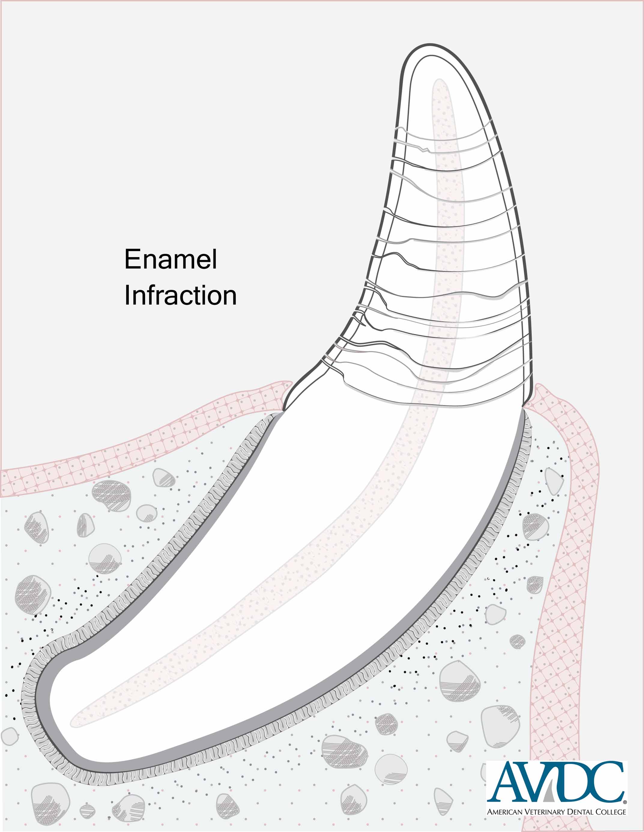
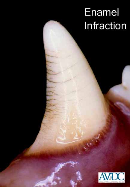
Enamel infraction (T/FX/EI):
Incomplete fracture (crack) of the enamel without loss of tooth substance
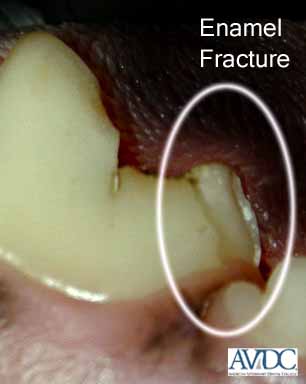
Enamel fracture (T/FX/EF):
Fracture with loss of crown substance confined to the enamel
Tooth Formation Abnormalities
_________________________________________
Persistent deciduous tooth (DT/P):
A deciduous tooth that is present when it should have exfoliated
Supernumerary tooth (T/SN):
Presence of an extra tooth (also called hyperdontia)
Hypodontia (HYP):
Developmental absence of few teeth
Oligodontia (OLI):
Developmental absence of numerous teeth
Anodontia (ANO):
Failure of all teeth to develop
Macrodontia (T/MAC):
Tooth/teeth are larger than normal
Microdontia (T/MIC):
Tooth/teeth are smaller than normal
Transposition (T/TRA):
Two teeth that have exchanged position
Fusion (T/FUS):
Combining of adjacent tooth germs and resulting in partial or complete union of the developing teeth; also called synodontia
Concrescence (T/CCR):
Fusion of the roots of two or more teeth at the cementum level
Fused roots (T/FDR):
Fusion of roots of the same tooth
Gemination (T/GEM):
A single tooth bud’s attempt to divide partially (cleft of the crown) or completely (presence of an identical supernumerary tooth); also called twinning
Supernumerary root (T/SR):
Presence of an extra root
Dilaceration (T/DIL):
Disturbance in tooth development, causing the crown or root to be abruptly bent or crooked
Dens invaginatus (T/DEN):
Invagination of the outer surface of a tooth into the interior, occurring in either the crown (involving the pulp chamber) or the root (involving the root canal); also called dens in dente
Enamel pearl (E/P):
Small, nodular growth on the root of a tooth made of enamel with or without a small dentin core and sometimes a covering of cementum
Unerupted tooth (T/U):
Tooth that has not perforated the oral mucosa
Embedded tooth (T/E):
Unerupted tooth covered in bone whose eruption is compromised by lack of eruptive force
Impacted tooth (T/I):
Unerupted or partially erupted tooth whose eruption is prevented by contact with a physical barrier
Dentigerous cyst (DTC):
Odontogenic cyst initially formed around the crown of a partially erupted or unerupted tooth; also called follicular cyst or tooth-containing cyst; removal is abbreviated DTC/R
Folliculitis (FOL):
Inflammation of the follicle of a developing tooth
Pericoronitis (PEC):
Inflammation of the soft tissues surrounding the crown of a partially erupted tooth
Tooth Resorption
_________________________________________
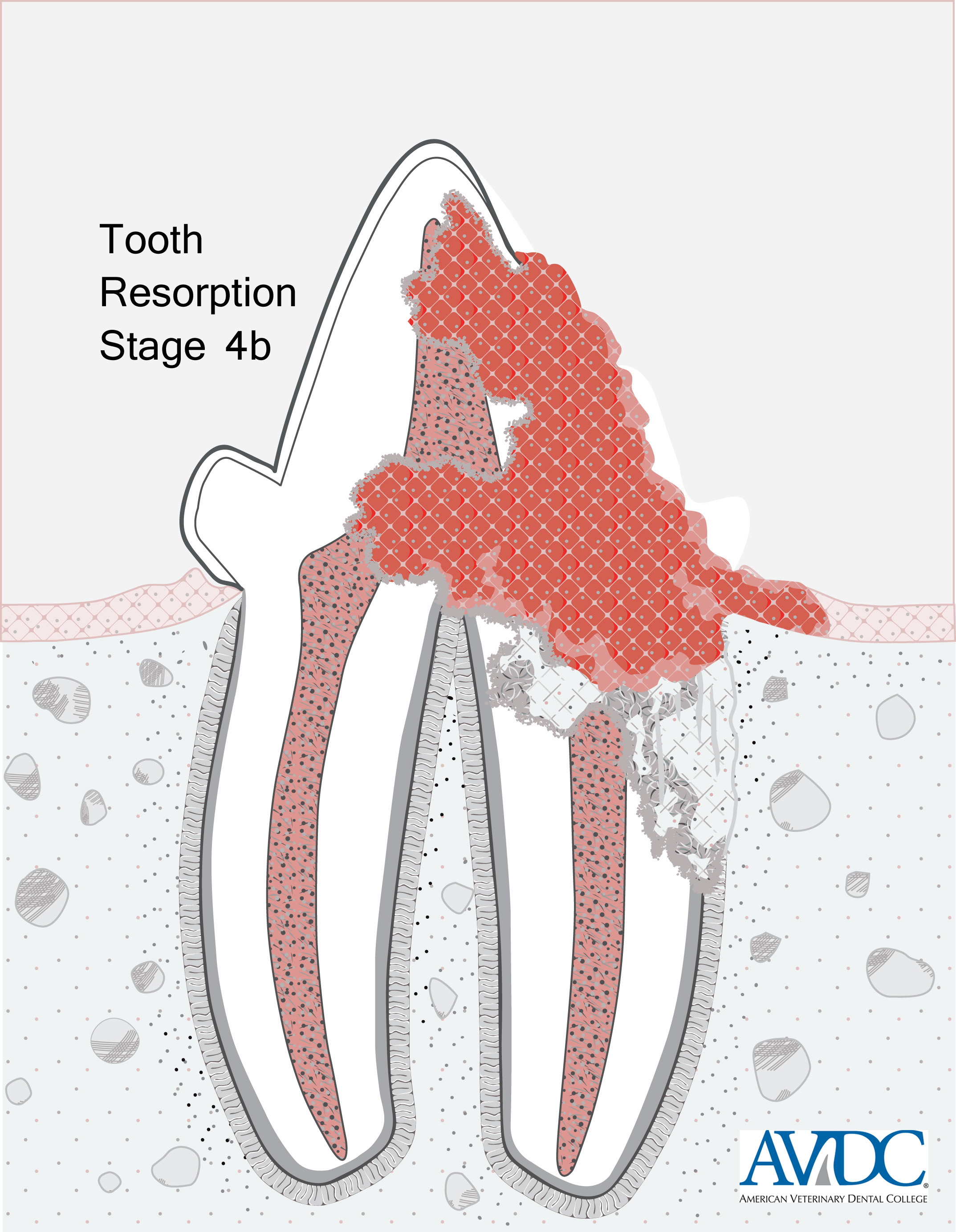
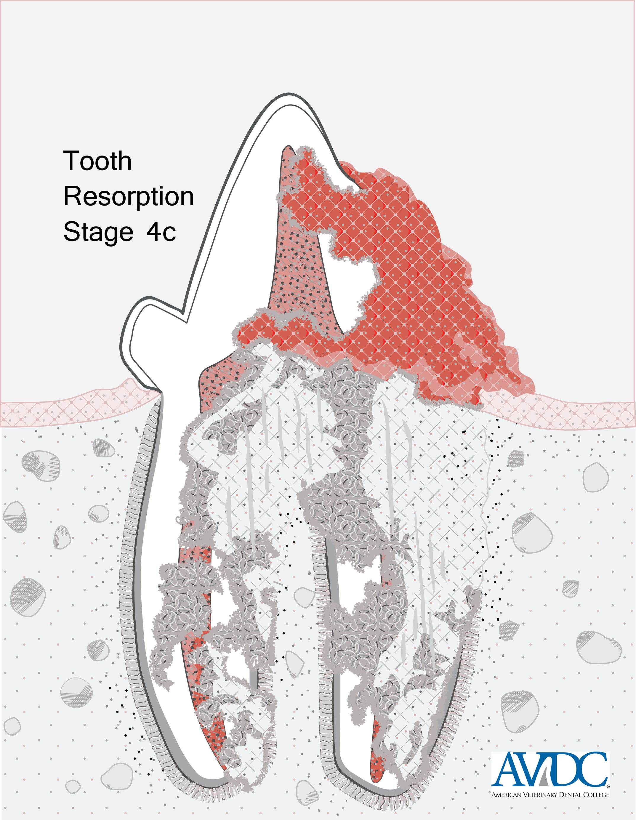
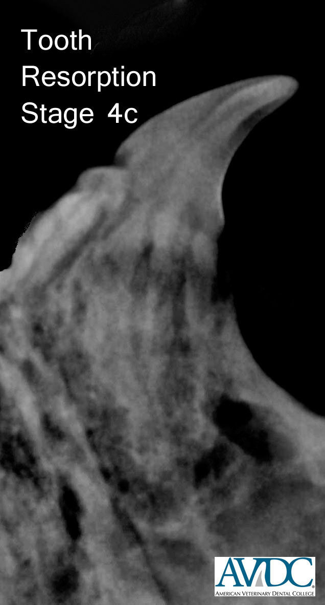
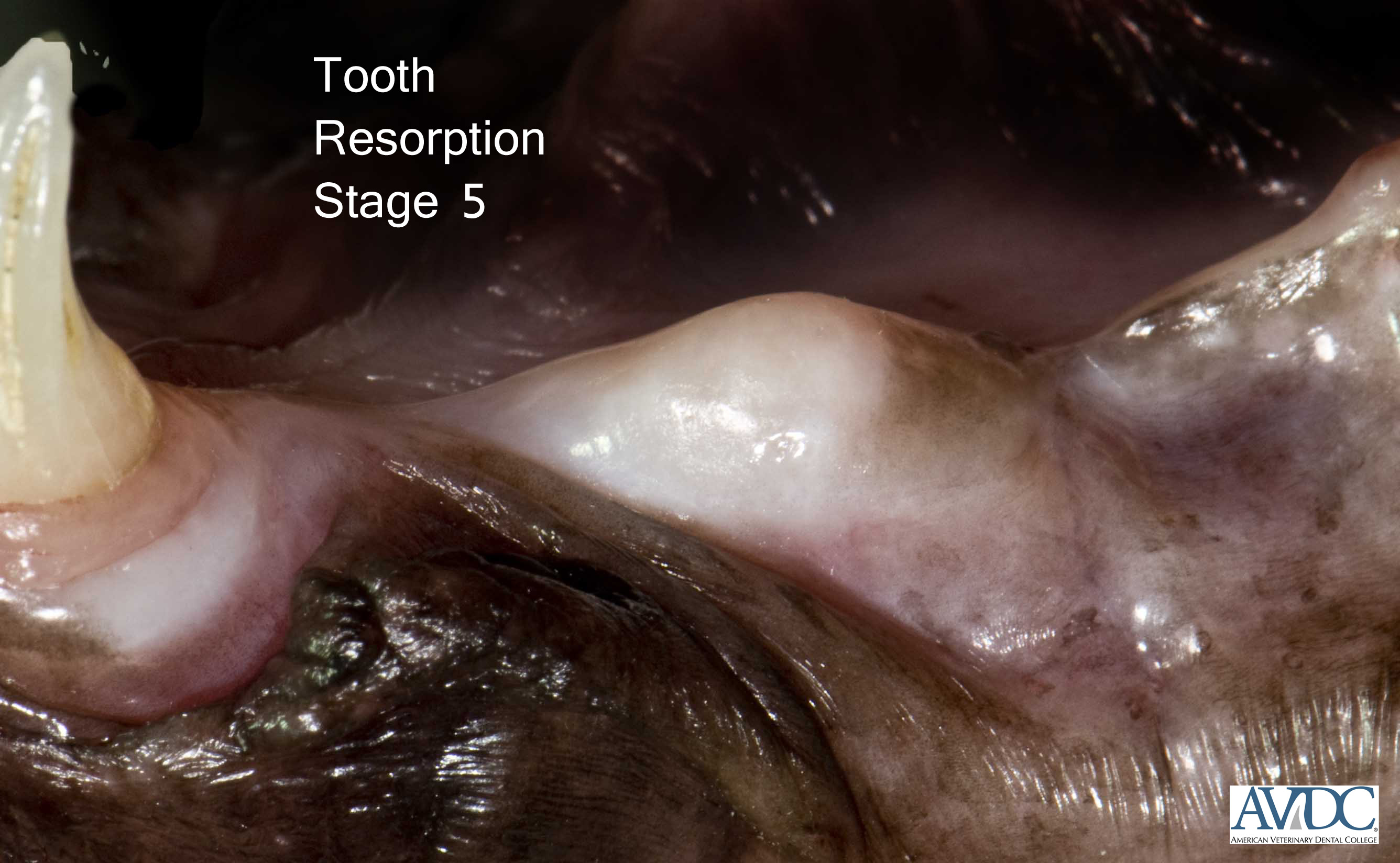
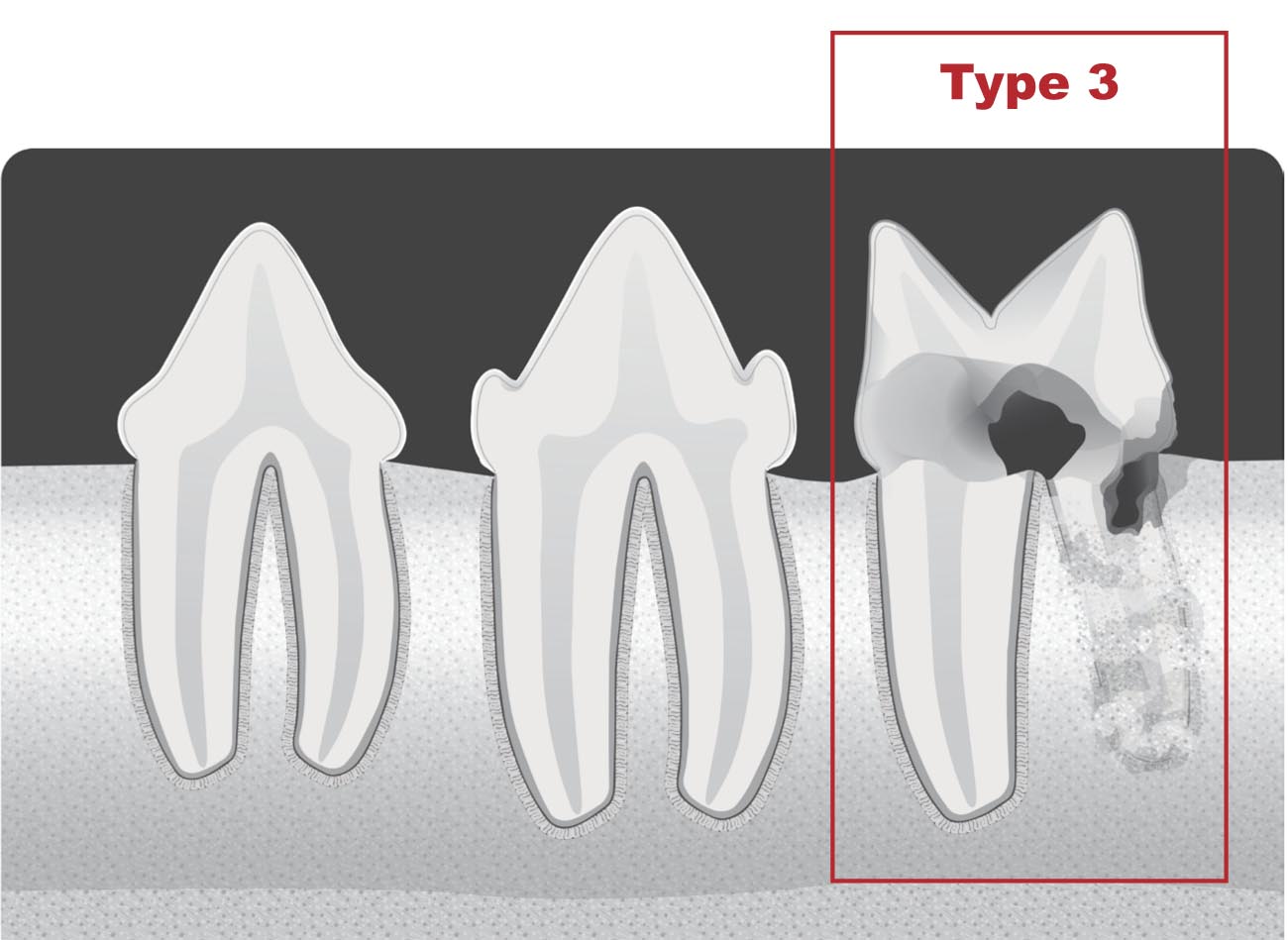
Tooth resorption is classified based on the severity of the resorption (Stages 1-5) and on the location of the resorption (Types 1-3).
The AVDC® classification of tooth resorption is based on the assumption that tooth resorption is a progressive condition.
Tooth resorption (TR):
Resorption of dental hard tissue
Internal resorption (RR:)
Tooth resorption originating within the pulp cavity
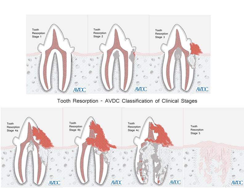
| Stage 1 (TR 1): Mild dental hard tissue loss (cementum or cementum and enamel). | 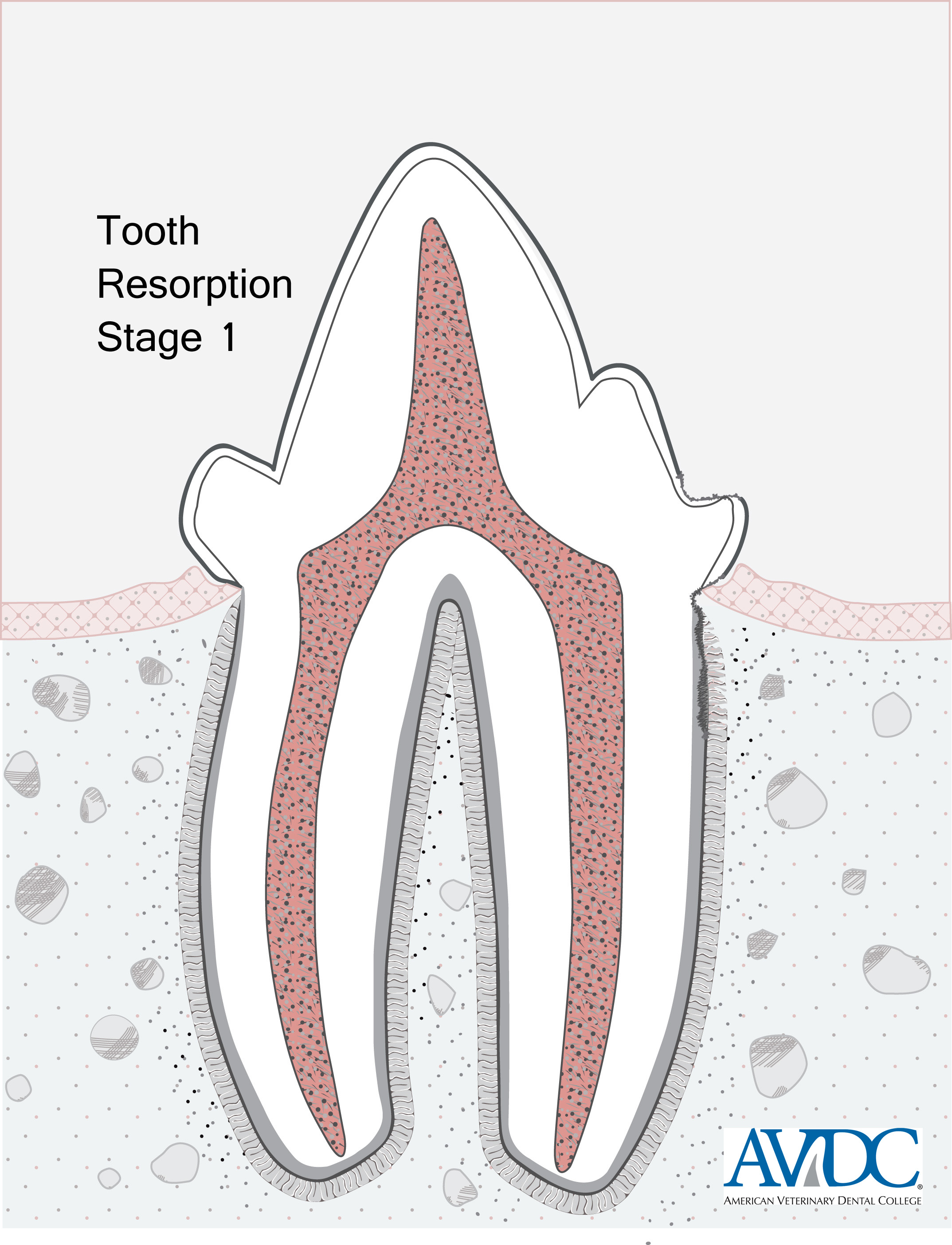 |
|
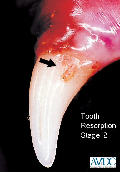
| Stage 2 (TR 2): Moderate dental hard tissue loss (cementum or cementum and enamel with loss of dentin that does not extend to the pulp cavity). | 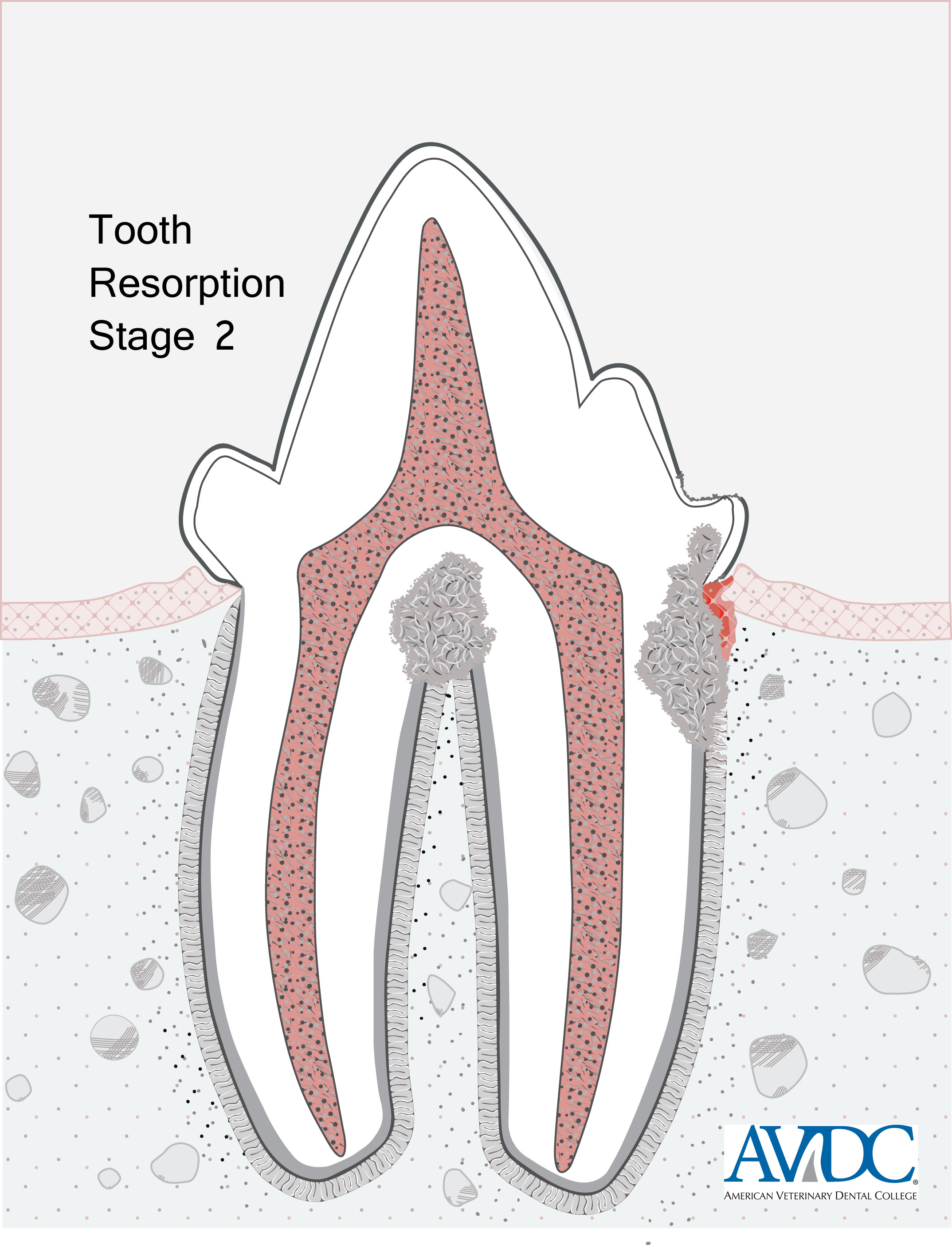 |
 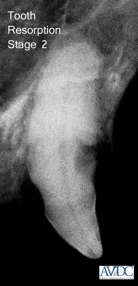 |
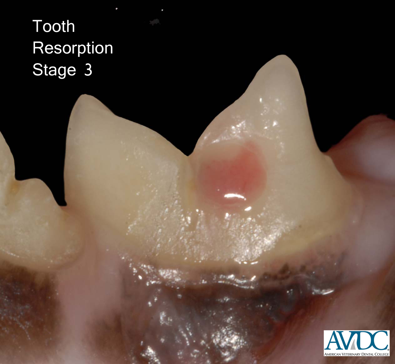
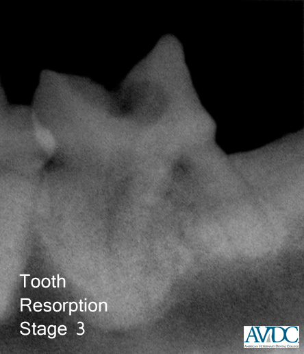
| Stage 3 (TR 3): Deep dental hard tissue loss (cementum or cementum and enamel with loss of dentin that extends to the pulp cavity); most of the tooth retains its integrity. | 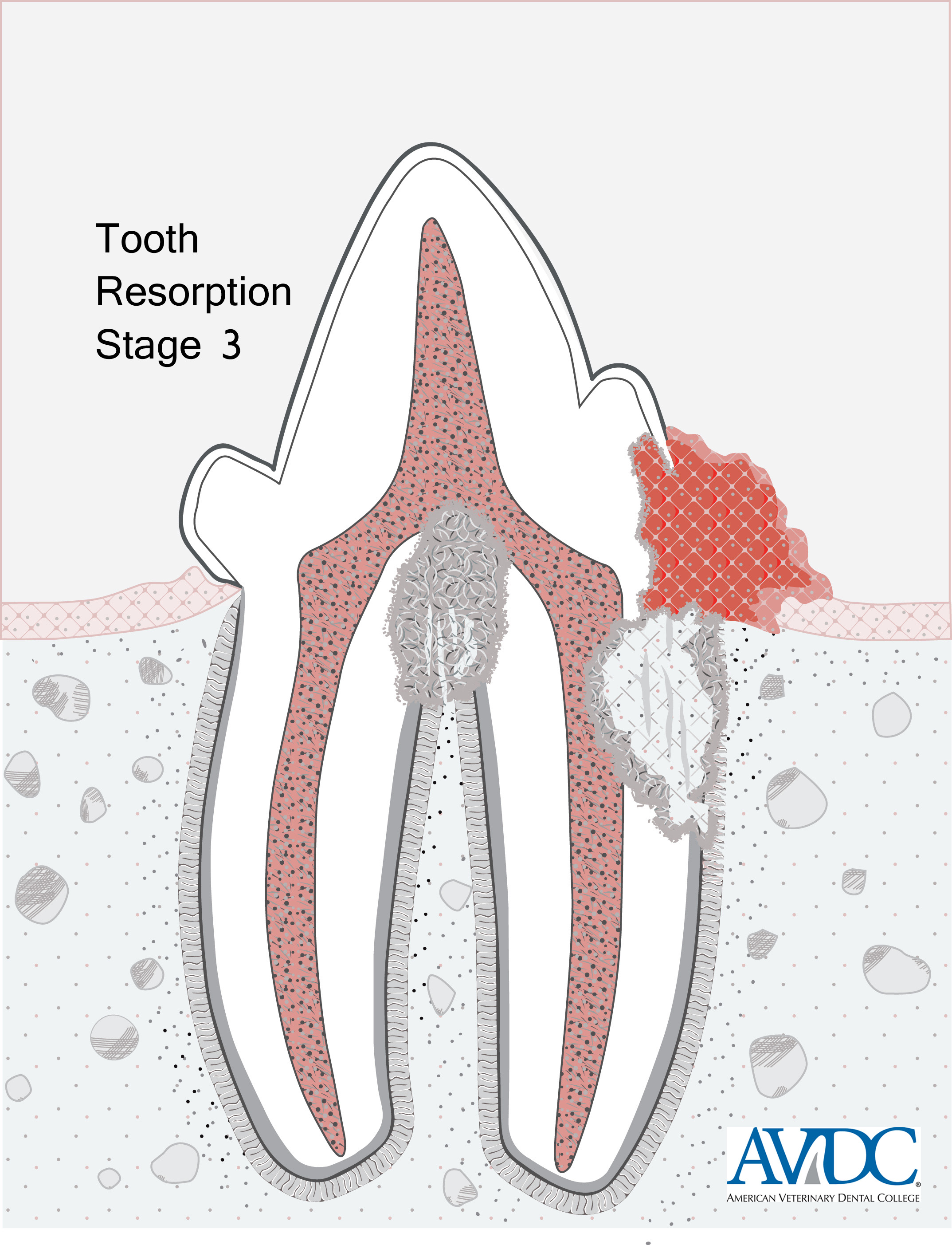 |
|
|
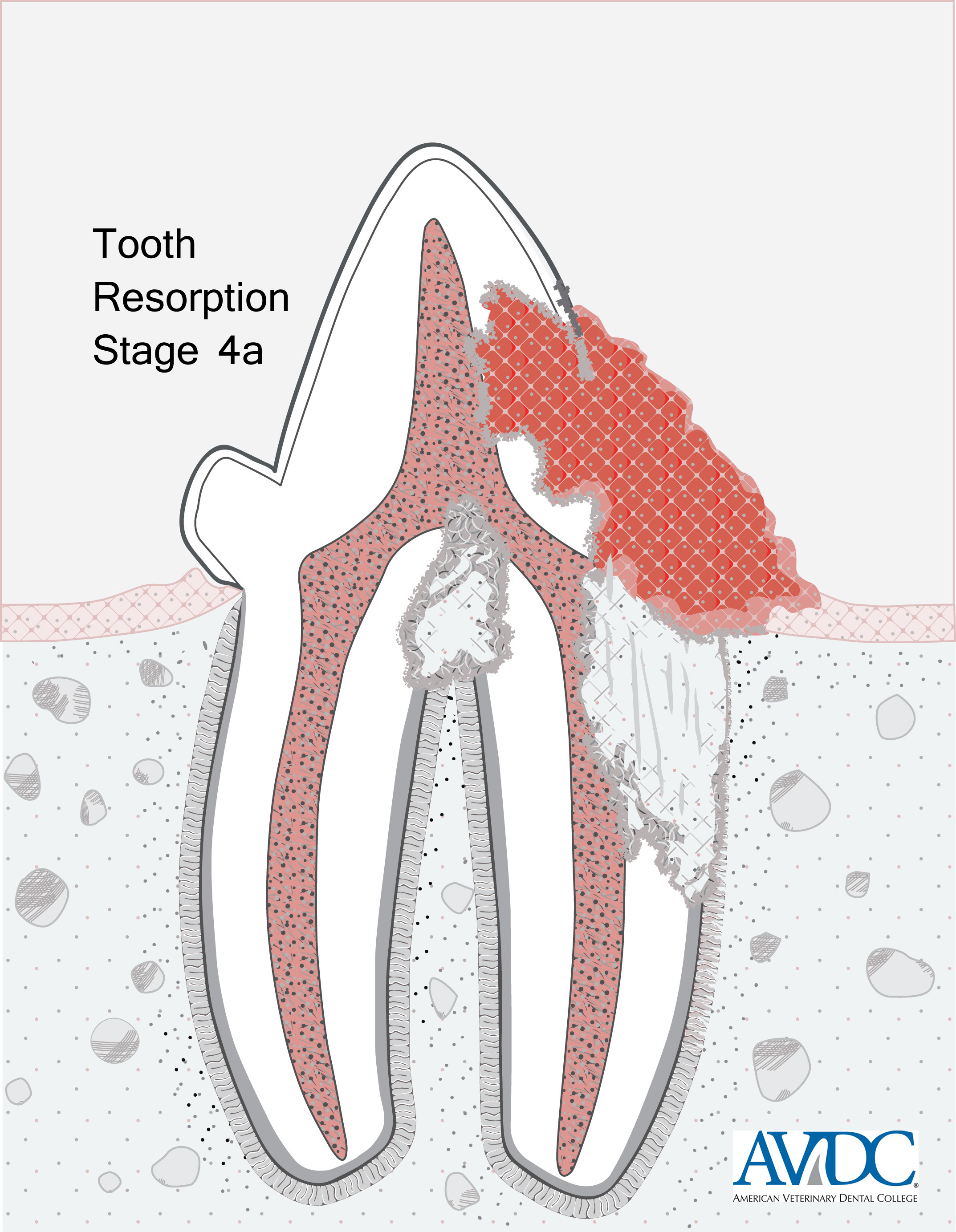
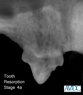
Stage 4 (TR 4): Extensive dental hard tissue loss (cementum or cementum and enamel with loss of dentin that extends to the pulp cavity); most of the tooth has lost its integrity. TR4a Crown and root are equally affected; |
 |
 |
|
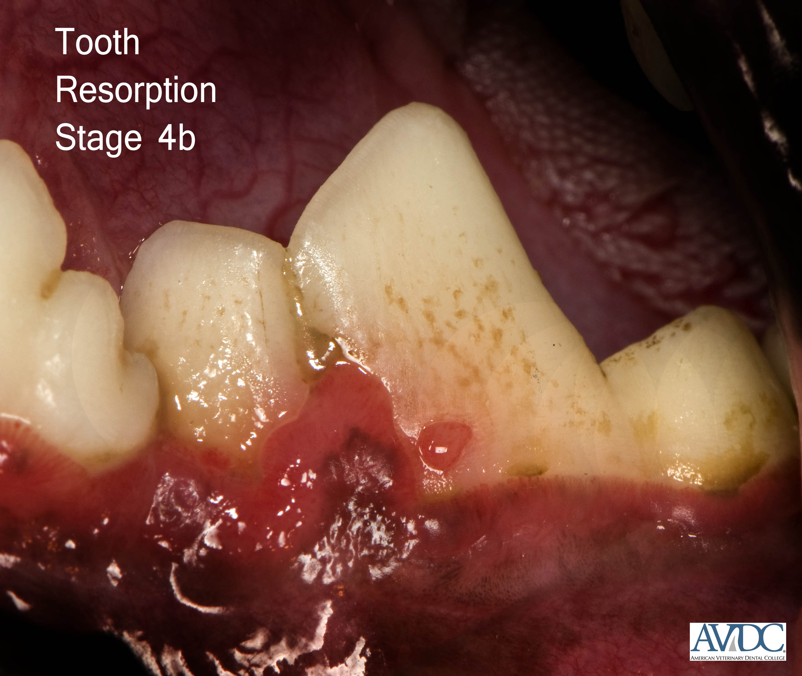
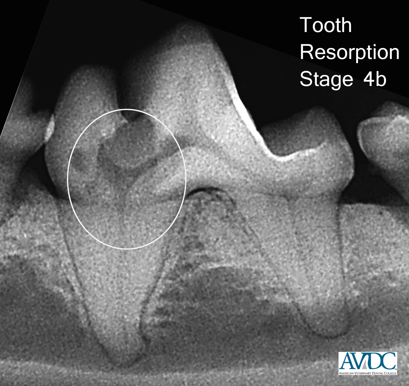
Stage 4 (TR 4): Extensive dental hard tissue loss (cementum or cementum and enamel with loss of dentin that extends to the pulp cavity); most of the tooth has lost its integrity. TR4b: Crown is more severely affected than the root; |
 |
|
|
Stage 4 (TR 4):: Extensive dental hard tissue loss (cementum or cementum and enamel with loss of dentin that extends to the pulp cavity); most of the tooth has lost its integrity. TR4c: Root is more severely affected than the crown. |
 |
  |
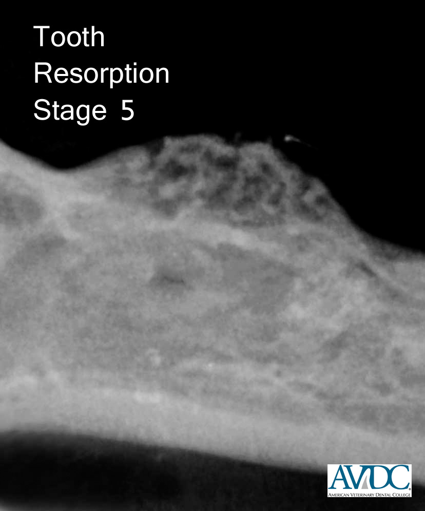
| Stage 5 (TR 5): Remnants of dental hard tissue are visible only as irregular radiopacities, and gingival covering is complete. | 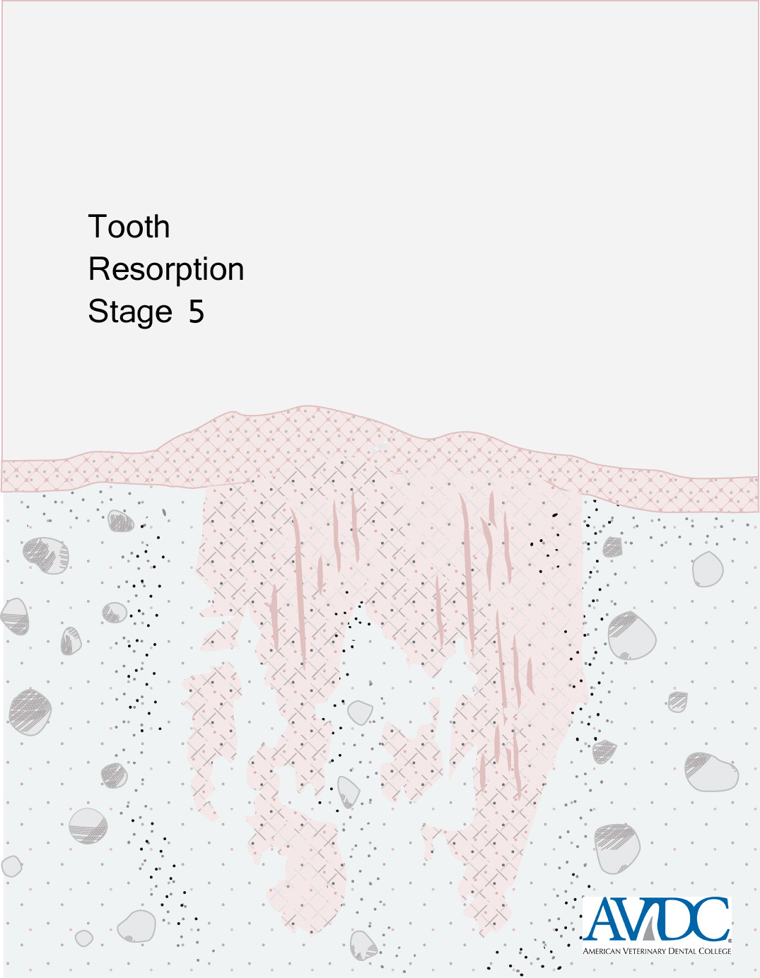 |
|
For low resolution printer-friendly versions of the full sets of tooth resorption images, click TR Diagrams or TR Clinical Images.
Types of Resorption Based on Radiographic Appearance
_________________________________________
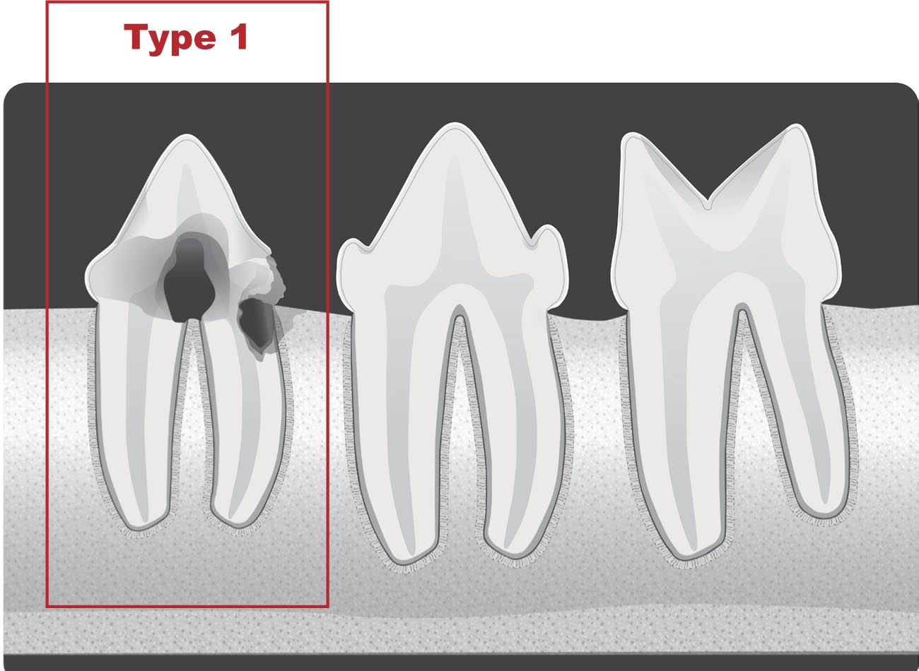
Type 1 (T1):
On a radiograph of a tooth with type 1 (T1) appearance, a focal or multifocal radiolucency is present in the tooth with otherwise normal radiopacity and normal periodontal ligament space.
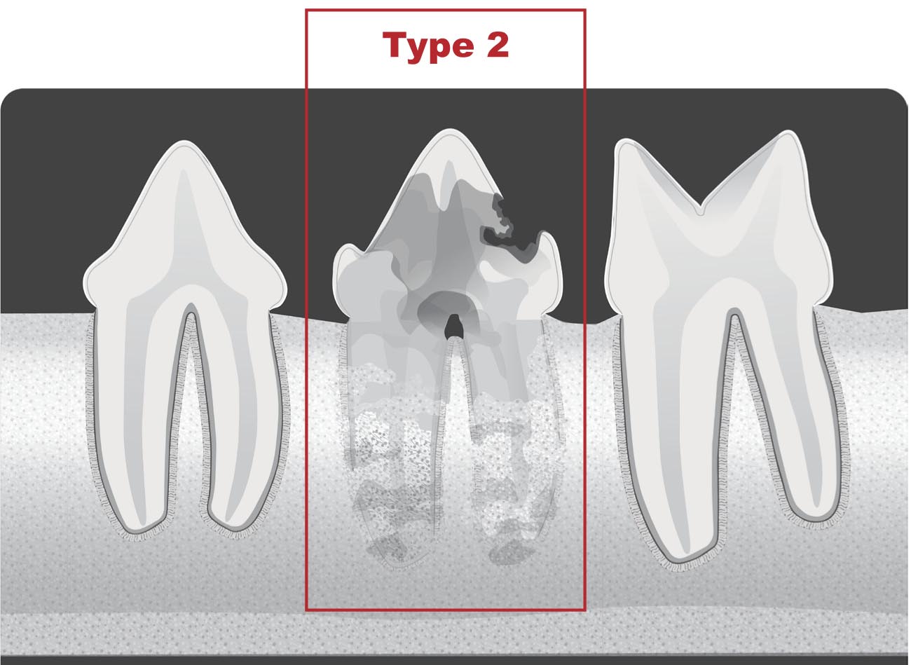
Type 2 (T2):
On a radiograph of a tooth with type 2 (T2) appearance, there is narrowing or disappearance of the periodontal ligament space in at least some areas and decreased radiopacity of part of the tooth.
Type 3 (T3):
On a radiograph of a tooth with type 3 (T3) appearance, features of both type 1 and type 2 are present in the same tooth. A tooth with this appearance has areas of normal and narrow or lost periodontal ligament space, and there is focal or multifocal radiolucency in the tooth and decreased radiopacity in other areas of the tooth.
Radiographic Examples of Types of Tooth Resorption:
![]()
![]()
![]()
Copyright of these images is owned by AVDC®. Download of these images and use in printed materials or presentations is permitted without charge provided that the source is cited as © AVDC® ®, used with permission. The diagrams are provided courtesy of Veterinary Information Network. The clinical images are provided by diplomates of AVDC®.
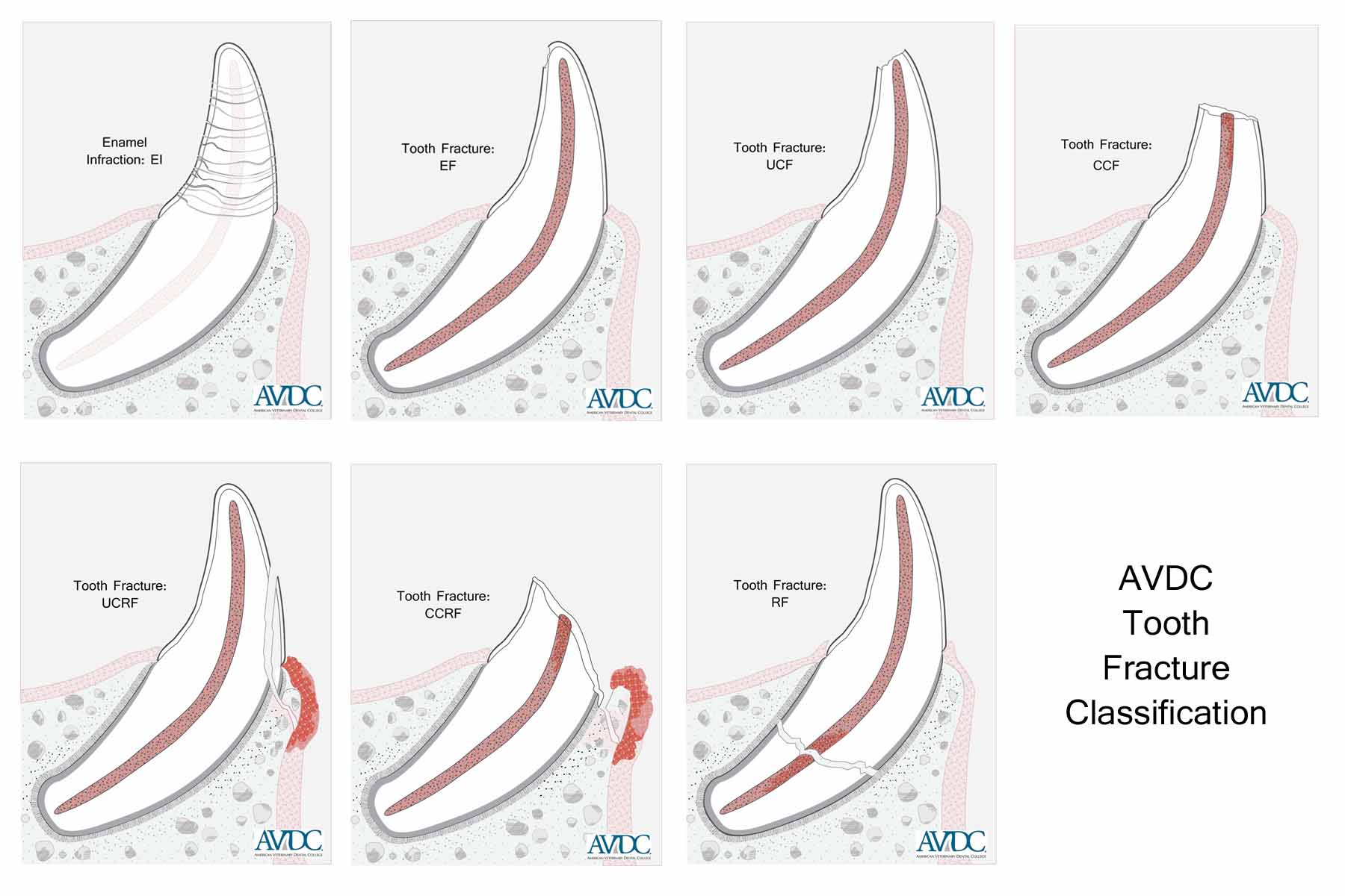
Tooth Fracture Classification
_________________________________________
The Tooth Fracture (T/FX) classification shown below can be applied for brachyodont and hypsodont teeth, which covers domesticated species and many wild species.
Fractures of teeth in some wild species may not fit into this classification because of differences in the tissues present in the teeth.
When used in AVDC® case log entries, the tooth fracture abbreviations noted below are to be stated as T/FX/{specific abbreviation} e.g T/FX/CCF
Enamel infraction (T/FX/EI):
Incomplete fracture (crack) of the enamel without loss of tooth substance
Enamel fracture (T/FX/EF):
Fracture with loss of crown substance confined to the enamel
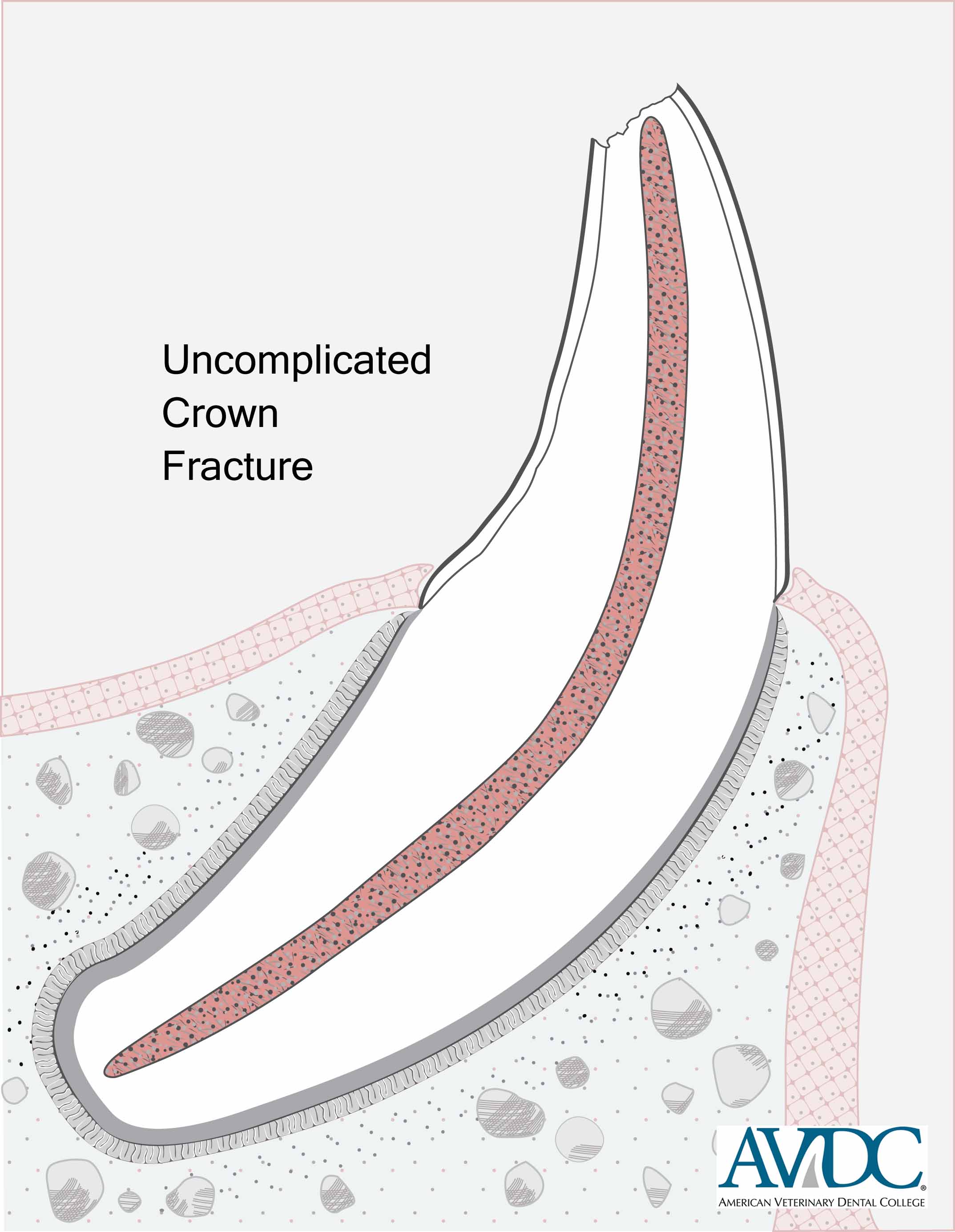
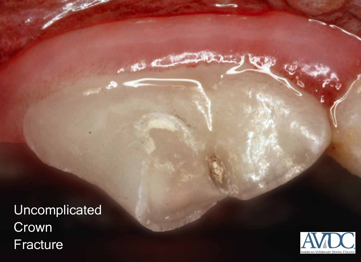
Uncomplicated crown fracture (T/FX/UCF):
Fracture of the crown that does not expose the pulp
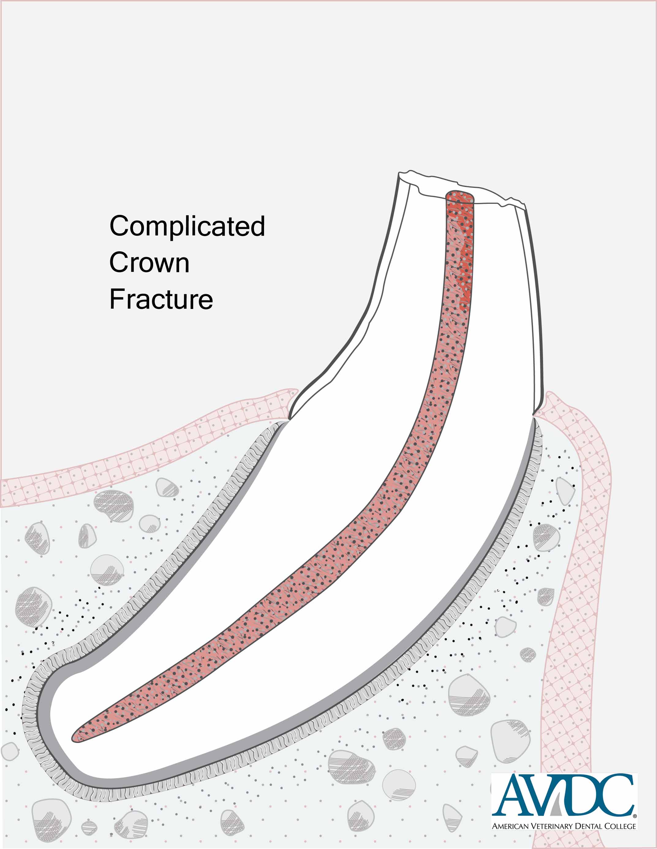
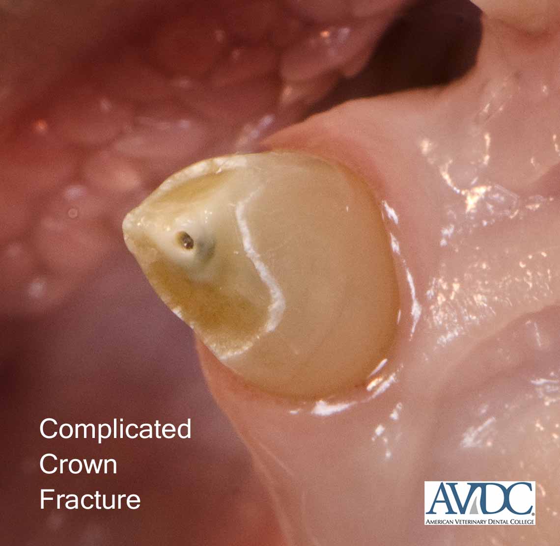
Complicated crown fracture (T/FX/CCF):
Fracture of the crown that exposes the pulp
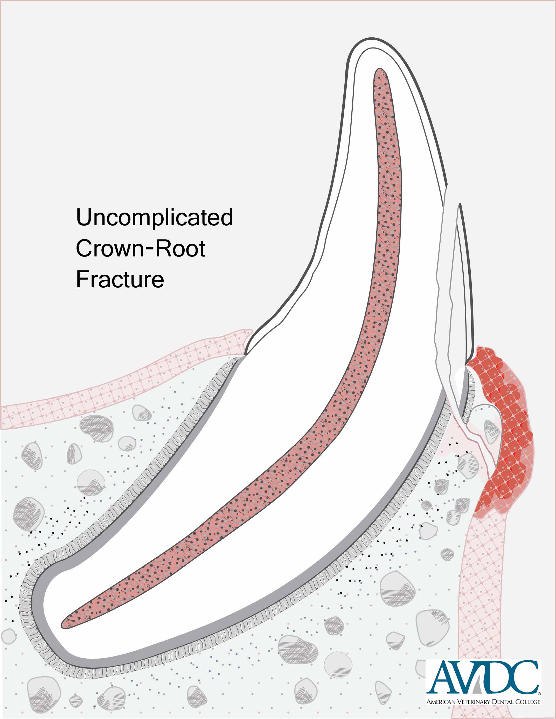
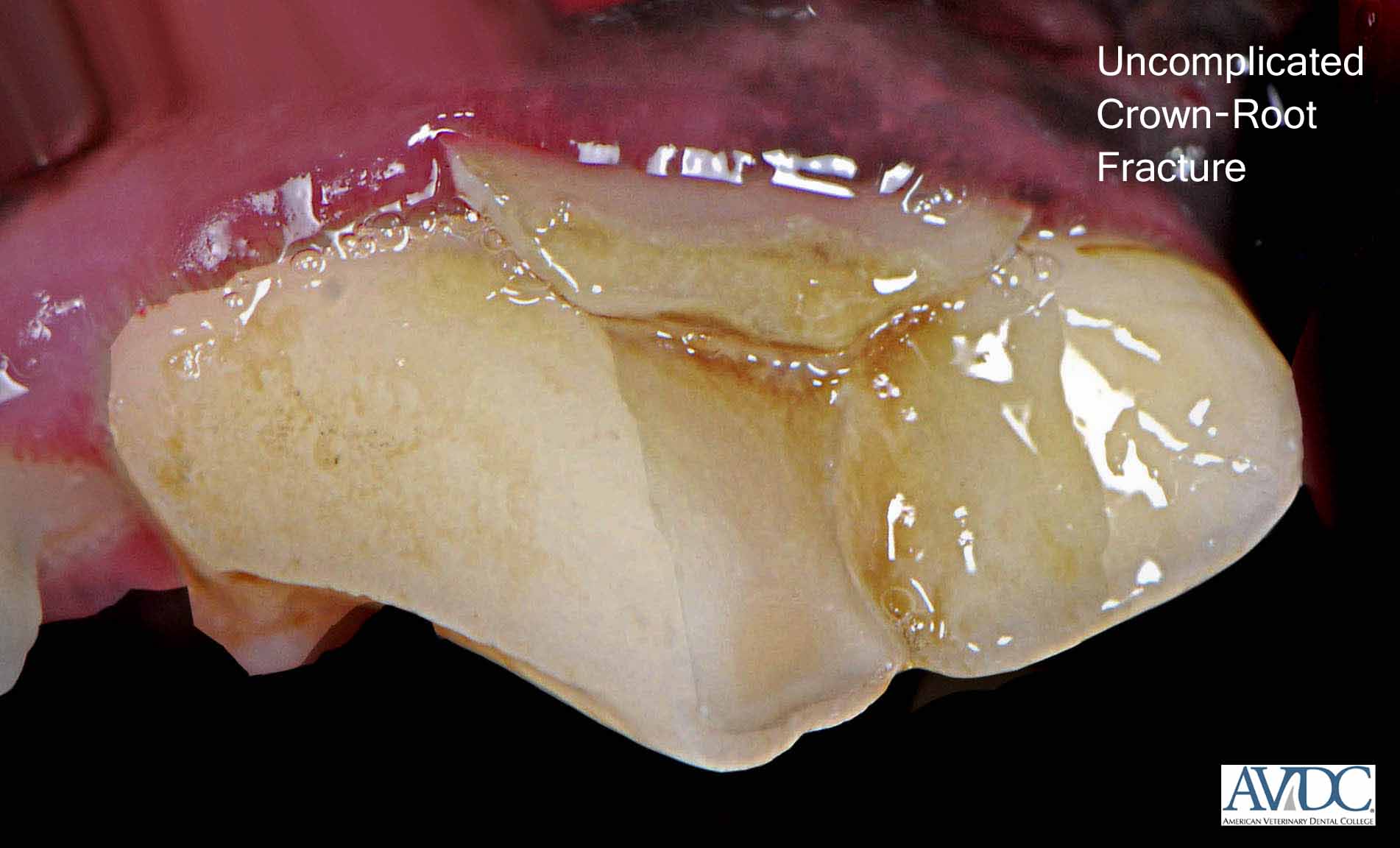
Uncomplicated crown-root fracture (T/FX/UCRF):
Fracture of the crown and root that does not expose the pulp
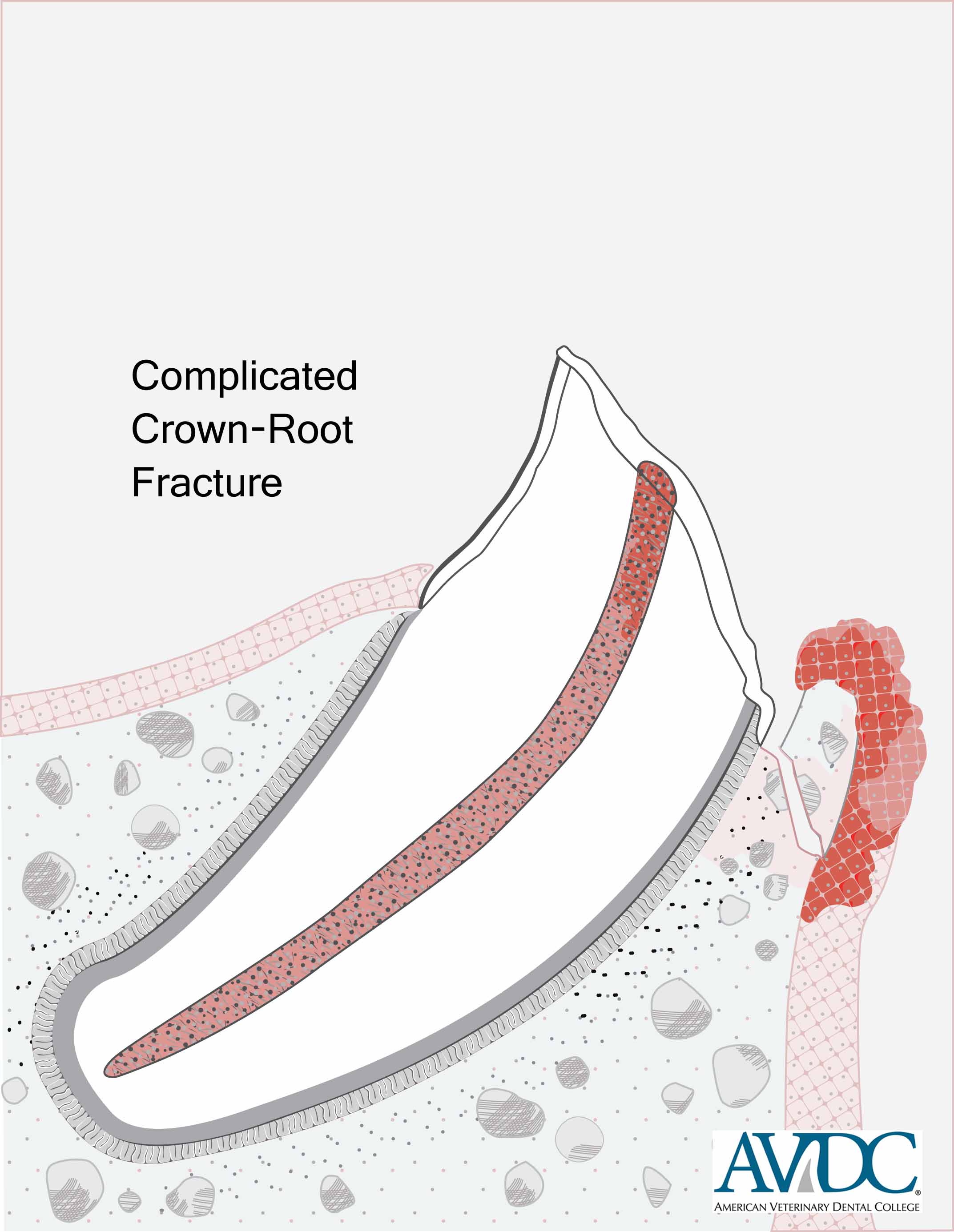
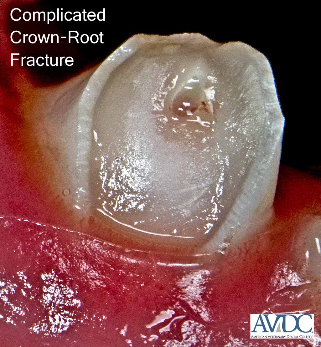
Complicated crown-root fracture (T/FX/CCRF):
Fracture of the crown and root that exposes the pulp
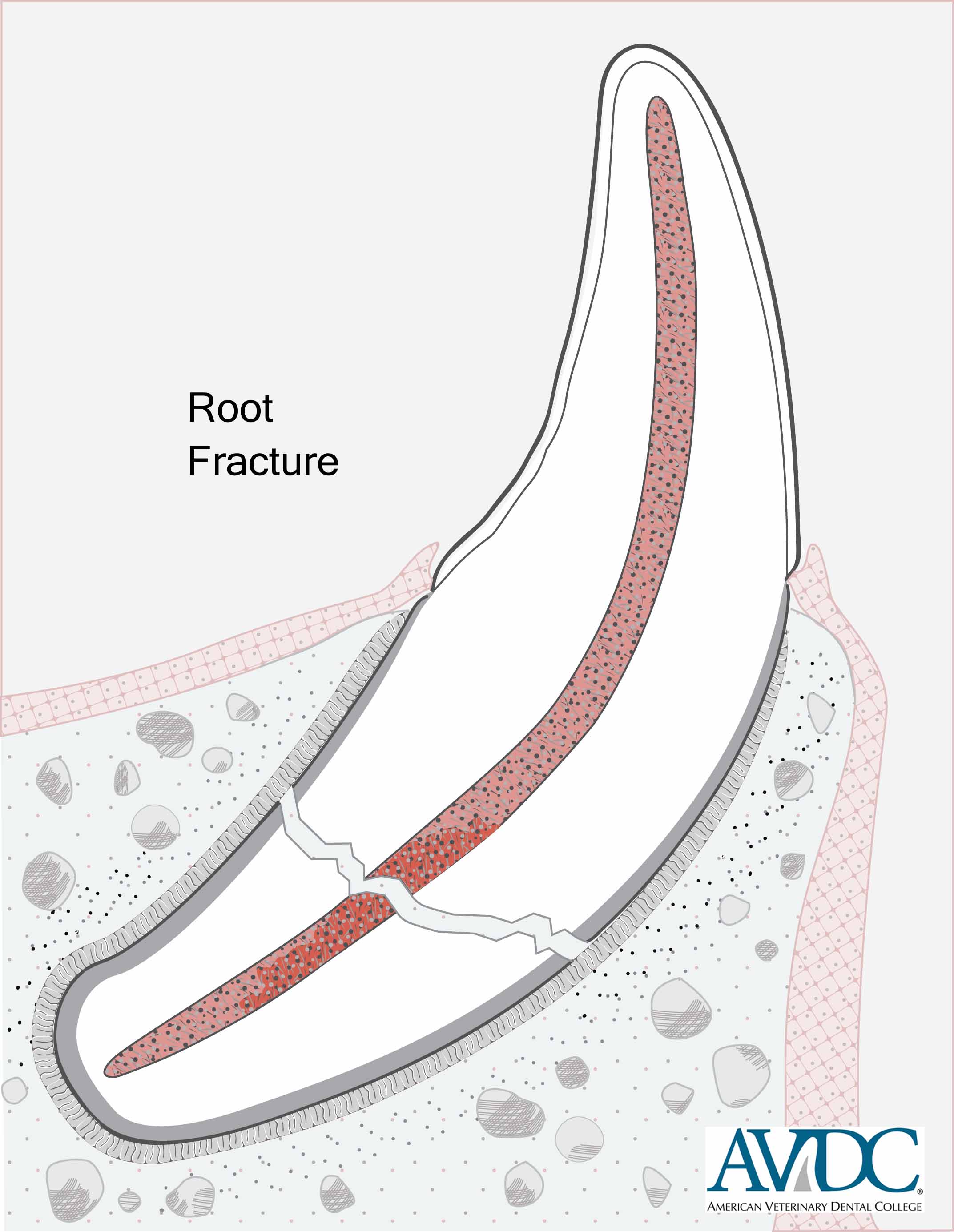
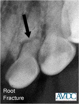
Root fracture (T/FX/RF):
Fracture involving the root
Retained root or reserve crown (RTR):
Presence of a root remnant or reserve crown remnant
Retained crown-root or clinical crown-reserve crown or clinical crown-reserve crown and root (RCR):
Presence of a crown-root remnant (in brachyodont teeth), clinical crown-reserve crown remnant (in aradicular hypsodont teeth) or clinical crown-reserve crown and root remnant (in radicular hypsodont teeth)
To download a .pdf printable version of this composite diagram, click download
Enamel infraction (EI):
An incomplete fracture (crack) of the enamel without loss of tooth substance. Example:
Enamel fracture (EF):
A fracture with loss of crown substance confined to the enamel. Example:

Uncomplicated crown fracture (UCF):
A fracture of the crown1 that does not expose the pulp. Example:
Complicated crown fracture (CCF):
A fracture of the crown1 that exposes the pulp. Example:
Uncomplicated crown-root fracture (UCRF):
A fracture of the crown and root that does not expose the pulp. Example:
Complicated crown-root fracture (CCRF):
A fracture of the crown and root that exposes the pulp. Example:
Root fracture (RF):
A fracture involving the root. Example:
Copyright of these images is owned by AVDC®. Download of the images and use in printed materials or presentations is permitted without charge provided that the source is cited as Copyright AVDC®, used with permission.
The AVDC® Board gratefully acknowledges Veterinary Information Network (VIN) for developing and donating the tooth fracture and tooth resorption diagrams. The clinical images are provided by diplomates of AVDC®.
To save a high-resolution version of an individual image for use in a presentation or a printed article, right-click on the image, click Save Picture As and follow the on-screen directions.
Endodontic Terminology
_________________________________________
Endodontics is a specialty in dentistry and oral surgery that is concerned with the prevention, diagnosis and treatment of diseases of the pulp-dentin complex and their impact on associated tissues.
Apexogenesis:
Physiological formation of the apex of a vital tooth
Pulp (PU):
Soft tissue in the pulp cavity
Odontoblasts:
Cells of mesenchymal origin that line the outer surface of the pulp and whose biological function is formation of dentin (dentinogenesis)
Predentin:
Unmineralized dentin matrix produced by odontoblasts
Dentin:
Mineralized tissue surrounding the pulp and containing dentinal tubules which radiate outward from the pulp to the periphery
Primary dentin:
Dentin produced until root formation is completed (e.g., dogs, cats) or the tooth comes into occlusion (e.g., horses)
Secondary dentin:
Dentin produced after root formation is completed
Tertiary dentin:
Dentin produced as a result of a local insult; can be reactionary (produced by existing odontoblasts) or reparative (produced by odontoblast-like cells that differentiated from pulpal stem cells as a result of an insult)
Sclerotic dentin:
Transparent dentin characterized by mineralization of the dentinal tubules as a result of an insult or normal aging
Periapical (PA):
Pertaining to tissues around the apex of a tooth, including the periodontal ligament and the alveolar bone
Fracture (FX):
Breaking of a bone or tooth
Vital tooth (T/V):
Tooth with vital pulp
Nonvital tooth (T/NV):
Tooth with nonvital pulp or from which the pulp has been removed
Pulp stones (PU/S):
Intrapulpal mineralized structures
Mineralization of the pulp (PU/M):
Pulpal mineralization resulting in regional narrowing or complete disappearance of the pulp cavity
Hypercementosis (HC):
Excessive deposition of cementum around the root or reserve crown of a tooth
Near pulp exposure (T/NE):
Thin layer of dentin separating the pulp from the outer tooth surface
Pulp exposure (T/PE):
Tooth with an opening through the wall of the pulp cavity uncovering the pulp
Tooth luxation (T/LUX):
Clinically or radiographically evident displacement of the tooth within its alveolus
Tooth avulsion (T/A):
Complete extrusive luxation with the tooth out of its alveolus
Periapical pathology (PA/P):
Pertaining to disease around the apex of a tooth
Periapical cyst (PA/C):
Odontogenic cyst formed around the apex of a tooth after stimulation and proliferation of epithelial rests in the periodontal ligament (also known as a radicular cyst)
Periapical granuloma (PA/G):
Chronic apical periodontitis with accumulation of mononuclear inflammatory cells and an encircling aggregation of fibroblasts and collagen that on diagnostic imaging appears as diffuse or circumscribed radiolucent lesion
Periapical abscess (PA/A):
Acute or chronic inflammation of the periapical tissues characterized by localized accumulation of suppuration
Osteosclerosis (OSS):
Excessive bone mineralization around the apex of a vital tooth caused by low-grade pulp irritation (asymptomatic; not requiring endodontic therapy)
Condensing osteitis (COO):
Excessive bone mineralization around the apex of a non-vital tooth caused by long-standing and low-toxic exudation from an infected pulp (requiring endodontic therapy)
Alveolar osteitis (AOS):
Inflammation of the alveolar bone considered to be a complication after tooth extraction
Osteomyelitis (OST):
Localized or wide-spread infection of the bone and bone marrow
Osteonecrosis (OSN):
Localized or wide-spread necrosis of the bone and bone marrow
Phoenix abscess:
Acute exacerbation of chronic apical periodontitis
Intraoral fistula (IOF):
Pathological communication between tooth, bone or soft tissue and the oral cavity; use IOF/R for its repair
Orofacial fistula (OFF):
Pathological communication between the oral cavity and face; use OFD/R for its repair
Indirect pulp capping (PCI):
Procedure involving the placement of a medicated material over an area of near pulp exposure
Direct pulp capping (PCD):
Procedure performed as part of vital pulp therapy and involving the placement of a medicated material over an area of pulp exposure
Vital pulp therapy (VPT):
Procedure performed on a vital tooth with pulp exposure, involving partial pulpectomy, direct pulp capping and access/fracture site restoration
Apexification (APN):
Procedure to promote apical closure of a nonvital tooth
Standard (orthograde) root canal therapy (RCT):
Procedure that involves accessing, debriding (including total pulpectomy), shaping, disinfecting, and obturating the root canal and restoring the access and/or fracture sites
Surgical (retrograde) root canal therapy (RCT/S):
Procedure that involves accessing the bone surface (through mucosa or skin), fenestration of the bone over the root apex, apicoectomy, and retrograde filling
Apicoectomy (AP/X):
Removal of the apex of a tooth; also called root end resection
Retrograde filling:
Restoration placed in the apical portion of the root canal after apicoectomy
Tooth repositioning (T/RP):
Repositioning of a displaced tooth
Interdental splinting (IDS):
Fixation using intraoral splints between teeth within a dental arch (for example for avulsed or luxated teeth that underwent reimplantation or repositioning); if performed for jaw fracture repair, use FX/R/IDS
Operative Dentistry and Prosthodontic Terminology
_________________________________________
Operative (or restorative) dentistry is a specialty in dentistry and oral surgery that is concerned with the art and science of the diagnosis, treatment and prognosis of defects of teeth that do not require prosthodontic crowns for correction.
Prosthodontics (or dental prosthetics or prosthetic dentistry) is a speciality in dentistry and oral surgery that is concerned with the provision of suitable substitutes for the clinical crown of teeth or for one or more missing or lost teeth and their associated parts. Maxillofacial prosthetics is considered a subspecialty of prosthodontics, involving palatal obturators and maxillofacial prostheses to replace resected or lost tissues.
Odontoplasty (ODY):
Surgical contouring of the tooth surface
Defect preparation (DP):
Removal of dental hard tissue to establish in a tooth the biomechanically acceptable form necessary to receive and retain a defect restoration
Restoration (R):
Anything that replaces lost tooth structure, teeth or oral tissues, including fillings, inlays, onlays, veneers, crowns, bridges, implants, dentures and obturators
Defect restoration:
Filling made of amalgam (R/A), glass ionomer (R/I), composite (R/C) or compomer (R/CP) within a prepared defect
Bridge (BRI):
Fixed partial denture used to replace a missing or lost tooth by joining permanently to adjacent teeth or implants
Crown preparation (CR/P):
Removal of enamel or enamel and dentin to establish on a tooth the biomechanically acceptable form necessary to receive and retain a prosthodontic crown
Temporary crown (CR/T):
Provisional, short-term cap made of resin to protect a prepared crown until cementation of a prosthodontic crown
Full crown:
Prosthodontic crown made of metal (CR/M), resin (CR/R), ceramic (CR/C) or porcelain fused to metal (CR/PFM) that covers the tip and all sides of a prepared crown
Partial crown:
Prosthodontic crown (e.g., three-quarter crown) made of metal (CR/M/P), resin (CR/R/P), ceramic (CR/C/P) or porcelain fused to metal (CR/PFM/P) that covers part of a prepared crown
Implant (IMP):
Titanium rod-shaped endosseous device to support intraoral prosthetics that resemble a tooth or group of teeth to replace one or more missing or lost teeth
Crown reduction (CR/XP):
Partial removal of tooth substance to reduce the height or an abnormal extension of the clinical crown
Crown amputation (CR/A):
Total removal of clinical crown substance
Post and core (PCB):
Placing a post into the root canal of a tooth that had root canal therapy and build-up of a core made of filling material around the portion of post that extends out from the pulp cavity
Anatomy of Jaws and TMJ
Clinically Relevant Terms Related to the Mandible and Temporomandibular Joint
Other Terms Relating to the Jaws and TMJ
Jaw Trauma and Management
Temporomandibular Joint Trauma and Other Conditions
——–
Jaw and TMJ Anatomy
_________________________________________
All mammals have two maxillas (or maxillae) and two mandibles. The adjective “maxillary” is often used in a wider sense, e.g., “maxillary fractures”, to include other facial bones, in addition to the maxillary bone proper.
References:
Anonymous. Nomina anatomica veterinaria. 4th ed. Zurich and Ithaca: World Association of Veterinary Anatomists, 1994. Evans HE. The skull. In: Evans HE, ed. Miller’s anatomy of the dog. 3rd ed. Philadelphia: W.B. Saunders, 1993;128-166. Hildebrand M. Analysis of vertebrate structure. 4th ed. New York: John Wiley & Sons, 1995. Nickel R, Schummer A, Seiferle E, et al. Teeth, general and comparative. In: The viscera of domestic mammals. 1st ed. Berlin: Verlag Paul Parey, 1973;75-99. Verstraete FJM. Maxillofacial fractures. In: Slatter DH, ed. Textbook of small animal surgery. 3rd ed. Philadelphia: WB Saunders Co, 2003;2190-2207.
Incisive Bones:
In domestic animals, the correct name for the paired bones that carry the maxillary incisors, located rostral to the maxillary bones, is the incisive bones, not the premaxilla.
Reference(s): Anonymous. Nomina anatomica veterinaria. 4th ed. Zurich and Ithaca: World Association of Veterinary Anatomists, 1994.
Clinically Relevant Terms Related to the Mandible and Temporomandibular Joint:
_________________________________________
Mandible
All animals have two mandibles, not one; removing one entire mandible is a total mandibulectomy not a hemimandibulectomy
Body of the mandible
The part that carries the teeth; often incorrectly referred to as horizontal ramus
Incisive part
The part that carries the incisors
Molar part
The part that carries the premolars and molars; premolar-molar part would probably have been more accurate
Alveolar margin
Often incorrectly referred to as alveolar crest
Ventral margin
Free ventral border
Mandibular canal
Contains a neurovascular bundle; often incorrectly referred to as the medullary cavity of the mandible
Mental foramens or foramina
Rostral, middle or caudal mental foramina in the dog and cat
Ramus of the mandible
The part that carries the 3 processes; often incorrectly referred to as the vertical ramus
Angular process
Caudoventral process (in carnivora)
Coronoid process
Process for the attachment of the temporal muscle
Condylar process
Consisting of mandibular head and mandibular neck; often incorrectly referred to as condyloid process
Mandibular head
Articular head of the condylar process
Mandibular neck
Neck of the condylar process
Mandibular notch
The notch on the caudal aspect, between the coronoid and condylar processes; not to be confused with the facial vascular notch
Mandibular angle
Angle between the body and ramus of the mandible.
Facial vascular notch
Shallow indentation on the ventral apsect of the mandible, rostral to the angular process (absent in carnivores)
Mandibular foramen
The entrance to the mandibular canal
Intermandibular joint (mandibular symphysis)
Median connection of the bodies of the right and left mandibles (in adult Sus and Equus replaced by a synostosis), consisting of intermandibular synchondrosis and intermandibular suture
Intermandibular synchondrosis
The smaller part of the intermandibular joint formed by cartilage
Intermandibular suture
The larger part of the intermandibular joint formed by connective tissue
Temporomandibular joint (TMJ)
The area where the condylar process of the mandible articulates with the mandibular fossa of the temporal bone
Articular disk
A flat structure composed of fibrocartilagenous tissue and positioned between the articular surfaces of the condylar process of the mandible and mandibular fossa of the temporal bone, separating the joint capsule in dorsal and ventral compartments; often incorrectly referred to as meniscus.
Mandibular fossa
Concave depression in the temporal bone that articulates with the mandibular head
Retroarticular process
A projection of the temporal bone that protrudes ventrally from the caudal end of the zygomatic arch and carries part of the mandibular fossa</
Reference: Anon. Nomina anatomica veterinaria. 4th ed. Zurich, Ithaca: World Association of Veterinary Anatomists, 1994.
Scapino RP. The third joint of the canine jaw. J Morphol 1965;116:23-50.
Other Terms Relating to the Jaws and TMJ
_________________________________________
Alveolar jugum (plural: alveolar juga):
The palpable convexity of the buccal alveolar bone overlying a large tooth root. References: Anonymous. Nomina Anatomica Veterinaria. 4th ed. Zurich and Ithaca: World Association of Veterinary Anatomists, 1994. Evans HE. Miller’s Anatomy of the Dog. 3rd ed. Philadelphia: WB Saunders Co, 1993.
Dental arch:
Referring to the curving structure formed by the teeth in their normal position; upper dental arch formed by the maxillary teeth, lower dental arch formed by the mandibular teeth
Jaw quadrant:
Referring to the left or right upper or lower jaw
Interarch:
Referring to between the upper and lower dental arches
Interquadrant:
Referring to between the left and right upper or lower jaw quadrants
Jaw and Related Abbeviations:
_________________________________________
Mandible/mandibular (MN):
Referring to the lower jaw
Maxilla/maxillary (MX):
Referring to the upper jaw
Mandibular symphysis (SYM):
Joint between the left and right mandibles (intermandibular joint)
Zygomatic arch (ZYG):
Consisting of the zygomatic process of the temporal bone and the temporal process of the zygomatic bone; also called zygoma
Jaw Trauma
_________________________________________
Maxillary fracture (MX/FX):
Fracture of the upper jaw (maxilla and other facial bones)
Mandibular fracture (MN/FX):
Fracture of the lower jaw (mandible)
Sympyseal separation (SYM/S):
Separation of the two mandibles in the mandibular symphysis; this includes parasymphyseal fractures where the fracture line is partly or completely paramedian to the symphysis; repair of symphyseal separation with wire (circumferential or interquadrant) and/or intraoral resin splinting is abbreviated with
Repair of a jaw fracture (FX/R):
Used when any of the other abbreviations do not describe the jaw fracture repair technique applied
Maxillomandibular fixation (FX/R/MMF):
Fixation that brings together the upper and lower jaws; use
MMF
for devices other than muzzles and splints
Muzzling (FX/R/MZ):
Maxillomandibular fixation using a prefabricated or custom-made muzzle; also used in horses to prevent eating (e.g., post sedation)
Interarch splinting (FX/R/IAS):
Maxillomandibular fixation using intraoral splints (commonly resin that can be reinforced with wire)
Interquadrant splinting (FX/R/IQS):
Fixation using intraoral splints (commonly resin that can be reinforced with wire) between the left and right upper or lower jaw quadrants
Interdental splinting (FX/R/IDS):
Fixation using intraoral splints (commonly resin that can be reinforced with wire) between teeth within a dental arch
Intraosseous wiring (FX/R/WIR/OS):
Fixation using intraosseous wire
Bone plating (FX/R/PL):
Fixation using bone plates
External skeletal fixation (FX/R/EXF):
Fixation using pins or wires and extraoral splinting
Wire cerclage (FX/R/WIR/C):
Fixation using circumferential wiring
Temporomandibular Joint Trauma and Other Conditions
_________________________________________
Decreased mouth opening (DMO):
Difficulty opening the mouth by the animal or decreased range of mouth opening upon oral examination
Temporomandibular joint fracture (TMJ/FX):
Fracture of one or more bony structures forming the temporomandibular joint; surgical repair is abbreviated with
TMJ/FX/R
Temporomandibular joint ankylosis (TMJ/A):
Fusion between the bones forming the temporomandibular joint or those in close proximity, resulting in progressive inability to open the mouth; removal of bone in ankylotic areas is abbreviated with
TMJ/A/R
Temporomandibular joint luxation (TMJ/LUX):
Displacement of the condylar process of the mandible; manual or surgical reduction of temporomandibular joint luxation is abbreviated with
TMJ/LUX/R
Temporomandibular joint dysplasia (TMJ/D):
Dysplasia of soft or hard tissues forming the temporomandibular joint
Open-mouth jaw locking (OMJL):
Inability to close the mouth due to locking of the coronoid process of the mandible ventrolateral to the ipsilateral zygomatic arch; manual reduction of open-mouth jaw locking is abbreviated with
OMJL/R
Zygomectomy (ZYG/X):
Resection (usually partial) of the zygomatic arch
Coronoidectomy (COR/X):
Resection (usually partial) of the coronoid process of the mandible
Condylectomy (CON/X):
Resection of the condylar process of the mandible
Definitions of Stage, Grade and Index
Periodontal Disease Stages
Furcation Involvement and Mobility Index
Tooth Mobility Index
Gingival and Periodontal Pathology
Periodontal Treatment
Flap Surgery
Flap Procedures
——–
Definitions of Stage, Grade and Index
_________________________________________
Stage:
The assessment of the extent of pathological lesions in the course of a disease that is likely to be progressive (e.g. stages of periodontal disease, staging of oral tumors).
Grade:
The quantitative assessment of the degree of severity of a disease or abnormal condition at the time of diagnosis, irrespective of whether the disease is progressive (e.g. a grade 2 mast cell tumor based on mitotic figures)
Index:
A quantitative expression of predefined diagnostic criteria whereby the presence and/or severity of pathological conditions are recorded by assessing a numerical value (e.g. gingival index, plaque index).
Stages of Periodontal Disease
_________________________________________
The degree of severity of periodontal disease (PD) relates to a single tooth; a patient may have teeth that have different stages of periodontal disease.
Normal (PD0):
Clinically normal; gingival inflammation or periodontitis is not clinically evident.
Stage 1 (PD1):
Gingivitis only without attachment loss; the height and architecture of the alveolar margin are normal.
Stage 2 (PD2):
Early periodontitis; less than 25% of attachment loss or, at most, there is a stage 1 furcation involvement in multirooted teeth. There are early radiologic signs of periodontitis. The loss of periodontal attachment is less than 25% as measured either by probing of the clinical attachment level, or radiographic determination of the distance of the alveolar margin from the cementoenamel junction relative to the length of the root.
Stage 3 (PD3):
Moderate periodontitis – 25-50% of attachment loss as measured either by probing of the clinical attachment level, radiographic determination of the distance of the alveolar margin from the cementoenamel junction relative to the length of the root, or there is a stage 2 furcation involvement in multirooted teeth.
Stage 4 (PD4):
Advanced periodontitis; more than 50% of attachment loss as measured either by probing of the clinical attachment level, or radiographic determination of the distance of the alveolar margin from the cementoenamel junction relative to the length of the root, or there is a stage 3 furcation involvement in multirooted teeth.
Reference: Wolf HF, Rateitschak EM, Rateitschak KH et al. Color atlas of dental medicine: periodontology, 3rd ed. Stuttgart: Georg Thieme Verlag, 2005.
Furcation Involvement and Mobility Index
_________________________________________
Furcation Index
Stage 1 (F1):
Furcation 1 involvement exists when a periodontal probe extends less than half way under the crown in any direction of a multirooted tooth with attachment loss.
Stage 2 (F2):
Furcation 2 involvement exists when a periodontal probe extends greater than half way under the crown of a multirooted tooth with attachment loss but not through and through.
Stage 3 (F3):
Furcation exposure exists when a periodontal probe extends under the crown of a multirooted tooth, through and through from one side of the furcation out the other.
Tooth Mobility Index
_________________________________________
Stage 0 (M0):
Physiologic mobility up to 0.2 mm.
Stage 1 (M1):
The mobility is increased in any direction other than axial over a distance of more than 0.2 mm and up to 0.5 mm.
Stage 2 (M2):
The mobility is increased in any direction other than axial over a distance of more than 0.5 mm and up to 1.0 mm.
Stage 3 (M3):
The mobility is increased in any direction than axial over a distance exceeding 1.0 mm or any axial movement.
Gingival and Periodontal Pathology
_________________________________________
Gingivitis:
Inflammation of gingiva
Periodontitis:
Inflammation of non-gingival periodontal tissues (i.e., the periodontal ligament and alveolar bone)
Gingival recession (GR):
Root surface exposure caused by apical migration of the gingival margin or loss of gingiva.
Gingival enlargement (GE):
Clinical term, referring to overgrowth or thickening of gingiva in the absence of a histological diagnosis
Gingival hyperplasia (GH):
Histological term, referring to an abnormal increase in the number of normal cells in a normal arrangement and resulting clinically in gingival enlargement
Abnormal tooth extrusion (ATE):
Increase in clinical crown length not related to gingival recession or lack of tooth wear
Alveolar bone expansion (ABE):
Thickening of alveolar bone at labial and buccal aspects of teeth
Periodontal Treatment
_________________________________________
Professional oral care
includes mechanical procedures performed in the oral cavity.
Professional dental cleaning (PRO)
refers to scaling (supragingival and subgingival plaque and calculus removal) and polishing of the teeth with power/hand instrumentation performed by a trained veterinary health care provider under general anesthesia. See also AVDC® Position Statements on Dental Health Care Providers and and on Non-Professional Dental Scaling.
Periodontal therapy
refers to treatment of diseased periodontal tissues that includes professional dental cleaning as defined above and one or more of the following: root planing, gingival curettage, periodontal flaps, regenerative surgery, gingivectomy/gingivoplasty, and local administration of antiseptics/antibiotics.
Home oral hygiene
refers to measures taken by pet owners that are aimed at controlling or preventing plaque and calculus accumulation.
Gingival curettage (GC):
Removal of damaged or diseased tissue from the soft tissue lining of a periodontal pocket.
Root planing (RP):
Removal of dental deposits from and smoothing of the root surface of a tooth; it is closed
(RP/C)
when performed without a flap or open
(RP/O)
when performed after creation of a flap.
Gingivectomy (GV):
Removal of some or all gingiva surrounding a tooth
Gingivoplasty (GV):
A form of gingivectomy performed to restore physiological contours of the gingiva
Guided Tissue Regeneration (GTR):
Regeneration of tissue directed by the physical presence and/or chemical activities of a biomaterial; often involves placement of barriers to exclude one or more cell types during healing of tissue
Crown lengthening (CR/L):
Increasing clinical crown height by means of gingivectomy/gingivoplasty, apically positioned flaps, post and core build-up, or orthodontic movement
Frenuloplasty (frenulotomy, frenulectomy) (FRE):
Reconstructive surgery or excision of a frenulum
Hemisection (HS):
Splitting of a tooth into two separate portionsB
Trisection (TS):
Splitting of a tooth into three separate portions.
Partial tooth resection (T/XP):
Removal of a crown-root segment with endodontic treatment of the remainder of the tooth
Root resection/amputation (RO/X):
Removal of a root with maintenance of the entire crown and endodontic treatment of the remainder of the tooth
Flap Surgery
_________________________________________
Flap (F): A sheet of tissue partially or totally detached to gain access to structures underneath or to be used in repairing defects; can be classified based on the location of the donor site (local or distant), attachment to donor site (pedicle, island or free), tissue to be transferred (e.g., mucosal, mucoperiosteal, cutaneous, myocutaneous), tissue thickness (partial-thickness or full-thickness), blood supply (random pattern or axial pattern), and direction and orientation of transfer (envelope, advancement, rotation, transposition, and hinged).
Location of Donor Site:
Local flap: Harvested from an adjacent site
Distant flap: Harvested from a remote site
Attachment to Donor Site:
Pedicle flap: Attached by tissue through which it receives its blood supply
Island flap (F/IS): Attached by a pedicle made up of only the nutrient vessels.
Free flap: Completely detached from the body; it has also been suggested that a free flap be termed a graft
Tissue to be Transferred:
Mucosal flap: Containing mucosa
Mucoperiosteal flap: Containing mucosa and underlying periosteum
Cutaneous (or skin) flap: Containing epidermis, dermis, and subcutaneous tissue
Myocutaneous flap: Containing skin and muscle
Gingival flap: Containing gingiva
Alveolar mucosa flap: Containing alveolar mucosa
Periodontal flap: Containing gingiva and alveolar mucosa
Labial flap: Containing lip mucosa
Buccal flap: Containing cheek mucosa
Sublingual flap: Containing sublingual mucosa
Palatal flap: Containing palatal mucosa
Pharyngeal flap: Containing pharyngeal mucosa
Tissue Thickness:
Partial-thickness (or split-thickness) flap: Consisting of a portion of the original tissue thickness
Full-thickness flap: Having the original tissue thickness
Blood Supply:
Random pattern flap: Randomly supplied by nonspecific arteries
Axial pattern flap: Supplied by a specific artery
Direction and Orientation of Transfer:
Envelope flap (F/EN): Retracted away from a horizontal incision; there is no vertical incision
Advancement (or sliding) flap (F/AD): Carried to its new position by a sliding technique in a direction away from its base
Rotation flap (F/RO): A pedicle flap that is rotated into a defect on a fulcrum point
Transposition flap (F/TR): Flap that combines the features of an advancement flap and a rotation flap
Hinged flap (F/HI): Folded on its pedicle as though the pedicle was a hinge; also called a turnover or overlapping flap
Apically positioned flap (F/AP): Moved apical to its original location
Coronally positioned flap (F/CO): Moved coronal to its original location
Mesiodistally or distomesially positioned flap: Moved distal or mesial to its original location along the dental arch; also called a laterally positioned flap (F/LA)
Oral Inflammation
Autoimmune Conditions Affecting the Mouth
Oral Tumors
Diagnostic and Non-Surgical Treatment Procedures
Surgical Treatment Procedures for Oral Tumors
Other Oral Pathology
——–
Oral Inflammation
_________________________________________
Note that a definitive diagnosis of inflammation often cannot be made based on physical examination findings alone.
Oral and oropharyngeal inflammation
is classified by location:
![]()
![]()
Gingivitis:
Inflammation of gingiva
Periodontitis:
Inflammation of non-gingival periodontal tissues (i.e. the periodontal ligament and alveolar bone)
Alveolar mucositis:
Inflammation of alveolar mucosa (i.e., mucosa overlying the alveolar process and extending from the mucogingival junction without obvious demarcation to the vestibular sulcus and to the floor of the mouth)
Sublingual mucositis:
Inflammation of mucosa on the floor of the mouth
Labial/buccal mucositis:
Inflammation of lip/cheek mucosa
![]()
![]()
Caudal mucositis:
Inflammation of mucosa of the caudal oral cavity, bordered medially by the palatoglossal folds and fauces, dorsally by the hard and soft palate, and rostrally by alveolar and buccal mucosa
Feline Stomatitis (FST):
A condition in the cat characterized by inflammation of the oral mucosa, often affecting the area immediately lateral to the palatoglossal folds (caudal stomatitis, ST/CS) with or without inflammation of other oral mucosa (i.e., gingiva, alveolar mucosa, labial/buccal mucosa, sublingual mucosa, and/or lingual mucosa); it commonly presents during the chronic stage, with or without (often proliferative) inflammation extending into mucosa of the oropharynx.
Contact mucositis and contact mucosal ulceration (CU):
Lesions in susceptible individuals that are secondary to mucosal contact with a tooth surface bearing the responsible irritant, allergen, or antigen. They have also been called “contact ulcers” and “kissing ulcers”.
![]()
![]()
Palatitis:
inflammation of mucosa covering the hard and/or soft palate
Glossitis:
inflammation of mucosa of the dorsal and/or ventral tongue surface
![]()
Osteomyelitis (OST): Inflammation of the bone and bone marrow
![]()
![]()
Cheilitis:
Inflammation of the lip (including the mucocutaneous junction area and skin of the lip)
Tonsilitis (TON/IN):
Inflammation of the palatine tonsil
Pharyngitis (PHA/IN):
inflammation of the pharynx
Autoimmune Conditions Affecting the Mouth
_________________________________________
Pemphigus vulgaris (PV):
Autoimmune disease characterized histologically by intraepithelial blister formation (after breakdown or loss of intercellular adhesion), biochemically by evidence of circulating autoantibodies against components of the epithelial desmosome-tonofilament complexes, and clinically by the presence of vesiculobullous and/or ulcerative oral and mucocutaneous lesions
Bullous pemphigoid (BUP):
Autoimmune disease characterized histologically by subepithelial clefting (separation at the epithelium-connective tissue interface), biochemically by evidence of circulating autoantibodies against components of the basement membrane, and clinically by the presence of erythematous, erosive, vesiculobullous and/or ulcerative oral lesions
Lupus erythematosis (LE):
Autoimmune disease characterized histologically by basal cell destruction, hyperkeratosis, epithelial atrophy, subepithelial and perivascular lymphocytic infiltration and vascular dilation with submucosal edema, biochemically by the evidence of circulating autoantibodies against various cellular antigens in both the nucleus and cytoplasm, and clinically by the presence of acute lesions (systemic LE) to skin, mucosa and multiple organs or chronic lesions (discoid LE) mostly confined to the skin of the face and mucosa of the oral cavity
Masticatory muscle myositis (MMM):
Autoimmune disease affecting the temporal, masseter, and medial and lateral pterygoid muscles of the dog. The term masticatory myositis is an acceptable alternative
Oral Tumors
_________________________________________
The AVDC® Nomenclature Committee is working with human oral pathologists, veterinary pathologists and veterinary oncologists to develop a set of names for specific tumor types that will be acceptable for standard use in veterinary dental patients.
The term “epulis” (plural = “epulides”) is a general term referring to a gingival mass lesion of any type. Examples of epulides include: focal fibrous hyperplasia, peripheral odotogenic fibroma, acanthomatous ameloblastoma, non-odontogenic tumors, pyogenic granulomas and reactive exostosis.
Types of Neoplasms Occurring in Oral Tissues (listed in alphabetical order)
Acanthomatous ameloblastoma (OM/AA):
A typically benign, but aggressive, histological variant of a group of epithelial odontogenic tumors known collectively as ameloblastomas which have a basic structure resembling the enamel organ (suggesting derivation from ameloblasts); the acanthomatous histological designation refers to the central cells within nests of odontogenic epithelium that are squamous and may be keratinized rather than stellate
Adenoma (OM/AD) :
Benign epithelial tumor in which the cells form recognizable glandular structures or in which the cells are derived from glandular epithelium
Adenocarcinoma (OM/ADC):
An invasive, malignant epithelial neoplasm derived from glandular tissue of either the oral cavity, nasal cavity or salivary tissue (major or accessory)
Amyloid producing odontogenic tumor (OM/APO):
A benign epithelial odontogenic tumor characterized by the presence of odontogenic epithelium and extra-cellular amyloid
Anaplastic neoplasm (OM/APN):
A malignant neoplasm whose cells are generally undifferentiated and pleomorphic (displaying variability in size, shape and pattern of cells and/or their nuclei)
Cementoma (OM/CE):
A benign odontogenic neoplasm of mesenchymal origin, consisting of cementum-like tissue deposited by cells resembling cementoblasts Biopsy
Feline inductive odontogenic tumor (OM/FIO):
A benign tumor unique to adolescent and young adult cats that originates multifocally within the supporting connective tissue as characteristic, spherical condensations of fibroblastic connective tissue associated with islands of odontogenic epithelium; has also been incorrectly called inductive fibroameloblastoma
Fibrosarcoma (OM/FS):
An invasive, malignant mesenchymal neoplasm of fibroblasts; a distinct histologically low-grade, biologically high-grade variant is often found in the oral cavity
Giant cell granuloma (OM/GCG):
A benign, tumor-like growth consisting of multi-nucleated giant cells within a background stroma on the gingiva (peripheral giant cell granuloma) or within bone (central giant cell granuloma); also called giant cell epulis
Granular cell tumor (OM/GCT):
A benign tumor of the skin or mucosa with uncertain histogenesis, most commonly occurring on the tongue; also called myoblastoma
Hemangiosarcoma (OM/HS):
A malignant neoplasm of vascular endothelial origin characterized by extensive metastasis; it has been reported in the gingiva, tongue and hard palate
Lipoma (OM/LI):
A benign mesenchymal neoplasm of lipocytes
Lymphosarcoma (OM/LS):
A malignant neoplasm defined by a proliferation of lymphocytes within solid organs such as the lymph nodes, tonsils, bone marrow, liver and spleen; the disease also may occur in the eye, skin, nasal cavity, oral cavity and gastrointestinal tract; also known as lymphoma
Malignant melanoma (OM/MM):
An invasive, malignant neoplasm of melanocytes or melanocyte precursors that may or may not be pigmented (amelanotic); also called melanosarcoma
Mast cell tumor (OM/MCT):
A local aggregation of mast cells forming a nodular tumor, having the potential to become malignant; also called mastocytoma
Multilobular tumor of bone (OM/MTB):
A potentially malignant and locally invasive neoplasm of bone that more commonly affects the mandible, hard palate and flat bones of the cranium with a multilobular histological pattern of bony or cartilaginous matrix surrounded by a thin layer of spindle cells that gives it a near pathognomonic radiographic “popcorn ball” appearance; also called multilobular osteochondrosarcoma, multilobular osteoma, multilobular chondroma, chondroma rodens, and multilobular osteosarcoma
Odontoma (OM/OD):
Odontogenic tumor that contains varying proportions of odontogenic epithelium, papillary ectomesenchyme, and dental hard matrices. It is rare in veterinary species, is typically found in younger animals, and generally manifests as unilateral and focal lesion. The compound odontoma (OM/ODD) is well-differentiated, often containing recognizable tooth-like structures (denticles) with enamel, dentin, cementum, and pulp, whereas the complex odontoma (OM/ODX) is less organized, usually containing a scrambled mix of odontogenic epithelium and dental hard matrices (mostly dentin or osteodentin) embedded in a fibrovascular stroma typically lacking overt features of ectomesenchyme of the dental papilla. Because the proliferative potential of the compound odontoma is limited, some investigators consider it to be a hamartomatous malformation (hamartoma) rather than a true neoplasm.
Osteoma (OM/OO):
A benign neoplasm of bone consisting of mature, compact, or cancellous bone
Osteosarcoma (OM/OS):
A locally aggressive malignant mesenchymal neoplasm of primitive bone cells that have the ability to produce osteoid or immature bone
Papilloma (OM/PAP):
An exophytic, pedunculated, cauliflower-like benign neoplasm of epithelium; canine papillomatosis is thought to be due to infection with canine papillomavirus in typically young dogs; severe papillomatosis may be recognized in older dogs that are immunocompromised
Peripheral nerve sheath tumor (OM/PNT):
A group of neural tumors arising from Schwann cells or perineural fibroblasts (or a combination of both cell types) of the cranial nerves, spinal nerve roots or peripheral nerves; they may be classified as histologically benign or malignant
Peripheral odontogenic fibroma (OM/POF):
A benign mesenchymal odontogenic tumor associated with the gingiva and believed to originate from the periodontal ligament; characterized by varying amounts of inactive-looking odontogenic epithelium embedded in a mature, fibrous stroma, which may undergo osseous metaplasia; historically been referred to as fibromatous epulis or – when bone or tooth-like hard tissue present within the lesion – ossifying epulis
Plasma cell tumor (OM/PCT):
A proliferation of plasma cells, commonly occurring on the gingiva or dorsum of the tongue; also called plasmacytoma
Rhabdomyosarcoma (OM/RBM):
A malignant neoplasm of skeletal muscle or embryonic mesenchymal cells”
Squamous cell carcinoma (OM/SCC):
An invasive, malignant epithelial neoplasm of the oral epithelium with varying degrees of squamous differentiation
Undifferentiated neoplasm (OM/UDN):
A malignant neoplasm whose cells are generally immature and lack distinctive features of a particular tissue type
Diagnostic and Non-Surgical Treatment Procedures
_________________________________________
Biopsy (B):
Removal of tissue from a living body for diagnostic purposes. The term has also been used to describe the tissue being submitted for evaluation
Guided biopsy:
Using computed tomography or ultrasonography to guide an instrument to the selected area for tissue removal
Surface biopsy (B/S):
Removal of tissue brushed, scraped or obtained by an impression smear from the intact or cut surface of a tissue in question
Needle aspiration (B/NA):
Removal of tissue by application of suction through a hollow needle attached to a syringe
Needle biopsy (B/NB):
Removal of tissue by puncture with a hollow needle
Core needle biopsy (B/CN):
Removal of tissue with a large hollow needle that extracts a core of tissue
Bite biopsy (B/B):
Removal of tissue by closing the opposing ends of an instrument
Punch biopsy (B/P):
Removal of tissue by a punch-type instrument
Incisional biopsy (B/I)
Removal of a selected portion of tissue by means of surgical cutting
Excisional biopsy (B/E):
Removal of the entire tissue in question by means of surgical cutting Guided biopsy – Using computed tomography or ultrasonography to guide an instrument to the selected area for tissue removal
Radiotherapy (RTH):
Use of ionizing radiation to control or kill tumor cells; also called radiation therapy
Chemotherapy (CTH):
Use of cytotoxic anti-neoplastic drugs (chemotherapeutic agents) to control or kill tumor cells
Immunotherapy (ITH):
Use of the immune system to control or kill tumor cells
Radiography (RAD):
Two-dimensional imaging of dental, periodontal, oral and maxillofacial structures using an X-ray machine and radiographic films, sensor pads or phosphor plates
Computed tomography (CT):
A method of medical imaging that uses computer-processed X-rays to produce tomographic images or ‘slices’ of specific areas of the body; digital geometry processing is used to generate three-dimensional images of an object of interest from a large series of two-dimensional X-ray images taken around a single axis of rotation
Cone-beam CT (CT/CB):
Variation of traditional CT that rotates around the patient, capturing data using a cone-shaped X-ray beam
Magnetic resonance imaging (MRI):
A method of medical imaging that uses the property of nuclear magnetic resonance to image nuclei of atoms inside the body
Ultrasonography (US):
A method of medical imaging of deep structures of the body by recording the echoes of pulses of ultrasonic waves directed into the tissues and reflected by tissue planes where there is a change in density
Scintigraphy (SCI):
A method of medical imaging that uses radioisotopes taken internally (e.g., by mouth, injection, inhalation), and the emitted radiation is captured by external detectors (gamma cameras) to form two-dimensional images
Surgical Treatment Procedures for Oral Tumors
_________________________________________
Surgery (S):
Branch of medicine that treats diseases, injuries and deformities by manual or operative methods
Buccotomy (BUC):
Incision through the cheek (for example to gain access to an intraoral procedure)
Cheiloplasty/commissuroplasty (CPL):
Reconstructive surgery of the lip/lip commissure
Commissurotomy (COM):
Incision through the lip commissure (for example to gain access to an intraoral procedure)
Partial mandibulectomy (S/M):
Surgical removal (en block) of part of the mandible and surrounding soft tissues
Dorsal marginal mandibulectomy (S/MD):
A form of partial mandibulectomy in which the ventral border of the mandible is maintained; also called marginal mandibulectomy or mandibular rim excision
Segmental mandibulectomy (S/MS):
A form of partial mandibulectomy in which a full dorsoventral segment of the mandible is removed
Bilateral partial mandibulectomy (S/MB):
Surgical removal of parts of the left and right mandibles and surrounding soft tissues
Total mandibulectomy (S/MT):
Surgical removal of one mandible and surrounding soft tissues
Partial maxillectomy (S/X):
Surgical removal (en block) of part of the maxilla and/or other facial bones and surrounding soft tissues
Bilateral partial maxillectomy (S/XB):
Surgical removal of parts of the left and right maxillae and/or other facial bones and surrounding soft tissues
Partial palatectomy (S/P):
Partial resection of the palate.
Other Oral Pathology
_________________________________________
Chewing lesion (CL):
Mucosal lesion resulting from self-induced bite trauma on the cheek (CL/B), lip (CL/L), palate (CL/P) or tongue/sublingual region (CL/T)
Eosinophilic granuloma (EOG):
Referring to conditions affecting the lip/labial mucosa (EOG/L), hard/soft palate (EOG/P), tongue/sublingual mucosa (EOG/T), and skin that are characterized histopathologically by the presence of an eosinophilic infiltrate
Pyogenic granuloma (PYO):
Inflammatory proliferation at the vestibular mucogingival tissues of the mandibular first molar tooth (in the cat probably due to malocclusion and secondary traumatic contact of these tissues by the ipsilateral maxillary fourth premolar tooth)
Erythema multiforme (EM):
Typically drug-induced hypersensitivity reaction characterized by erythematous, vesiculobullous and/or ulcerative oral and skin lesions
Calcinosis circumsctripta (CC):
Circumscribed areas of mineralization characterized by deposition of calcium salts (e.g., in the tip of the tongue)
Retrobulbar abscess (RBA):
Abscess behind the globe of the eye
Retropharyngeal abscess (RPA):
Abscess behind the pharynx
Craniomandibular osteopathy (CMO):
Disease characterized by cyclical resorption of normal bone and excessive replacement by immature bone along mandibular, temporal and other bone surfaces in immature and adolescent dogs
Calvarial hyperostosis (CHO):
Disease characterized by irregular, progressive proliferation and thickening of the cortex of the bones forming the calvarium in adolescent dogs
Fibrous osteodystrophy (FOD):
Disease characterized by the formation of hyperostotic bone lesions, in which deposition of unmineralized osteoid by hyperplastic osteoblasts and production of fibrous connective tissue exceed the rate of bone resorption; usually due to primary or secondary hyperparathyroidism; resulting in softened, pliable and distorted bones of the face (“rubber jaw”, “bighead” or “bran disease”)
Periostitis ossificans (PEO):
Periosteal new bone formation in immature dogs, manifesting clinically as (usually) unilateral swelling of the mid to caudal body of the mandible and radiographically as two-layered (double) ventral mandibular cortex
Anatomy of Tongue, Lips, Cheek and Palate
Abnormalities of the Palate
Anatomy of the Nose, Pharynx, Tonsils and Face
Nasal, Facial and Pharyngeal Abnormalities
Anatomy of the Salivary Gland Glands
Salivary Gland Abnormalities
Lymph Nodes
——–
Anatomy of the Tongue, Lips, Cheek and Palate
_________________________________________
Tongue (LIN):
Fleshy muscular organ in the mouth used for tasting, licking, swallowing, articulating and thermoregulation; use
LIN/X
for tongue resection
Lip/cheek (LIP):
Fleshy parts that form the upper and lower edges of the opening of the mouth/side of the face below the eye; use
LIP/X
for lip/cheek resection
LIP/A
Lip avulsion (LIP/A): A traumatic separation of the lip from the underlying connective tissue; use LIP/A/R for lip avulsion repair
Hard palate:
The part of the palate supported by bone.
The midline of the hard palate is not a symphysis but is formed by the interincisive suture, the median palatine suture of the palatine processes of the maxillary bones, and the median suture of the palatine bones.
References: Anonymous. Nomina anatomica veterinaria. 4th ed. Zurich and Ithaca: World Association of Veterinary Anatomists, 1994. Evans HE. Miller’s anatomy of the dog. 3rd ed. Philadelphia: WB Saunders Co, 1993.
Palatine rugae:
Transverse ridges of mucosa on the hard palate
Incisive papilla:
Elevation of mucosa at the rostral end of the median line of junction of the halves of the palate, concealing the orifices of the incisive ducts
Soft palate:
The caudal part of palate that is not supported by bone
Abnormalities of the Palate
_________________________________________
Palate defect (PDE):
Acquired communication between the oral and nasal cavities along the hard or soft palate; surgical repair is abbreviated with
PDE/R
Cleft lip (CFL):
Congenital longitudinal defect of the upper lip or upper lip and most rostral hard palate (regardless of location); surgical repair is abbreviated with
CFL/R
Cleft palate (CFP):
Congenital longitudinal defect in the midline of the hard and soft palate; surgical repair is abbreviated with
CFP/R
Cleft soft palate (CFS):
Congenital longitudinal defect in the midline of the soft palate only; surgical repair is abbreviated with
CFS/R
Unilateral soft palate defect (CFSU):
Congenital longitudinal defect of the soft palate on one side only; surgical repair is abbreviated with
CFSU/R
Soft palate hypoplasia (CFSH):
Congenital decrease in length of the soft palate; surgical lengthening of the soft palate is abbreviated with
CFSH/R
Traumatic cleft palate (CFT):
Acquired longitudinal defect in the midline of the hard and/or soft palate resulting from trauma; surgical repair is abbreviated with
CFT/R
Oronasal fistula (ONF):
Acquired communication between the oral and nasal cavities along the upper dental arch; surgical repair is abbreviated with
ONF/R
Oroantral fistula (OAF):
Acquired communication between the oral cavity and maxillary sinus in pigs, ruminants and equines (also called oromaxillary fistula in equines); surgical repair is abbreviated with
OAF/R
Elongated soft palate (ESP):
Congenital increase in length of the soft palate; surgical reduction of the soft palate is abbreviated with
ESP/R
Palatal obturator (POB):
Prosthetic device for temporary or permanent closure of palate defects
Anatomy of the Nose, Pharynx, Tonsil and Face
_________________________________________
Palatine tonsil (TON):
Tonsil related to the lateral attachment of the soft palate
Tonsillar fossa:
Depression containing the palatine tonsil
Semilunar fold:
Mucosal fold from the ventrolateral aspect of the soft palate, forming the medial wall of the tonsillar fossa
Pharynx (PHA):
Throat caudal to the oral cavity and divided into nasopharynx and oropharynx
Fauces:
The fauces are defined as the lateral walls of the oropharynx that are located medial to the palatoglossal folds. The areas lateral to the palatoglossal fold, commonly involved in feline stomatitis, are not the fauces.
References: Anonymous. Nomina anatomica veterinaria. 4th ed. Zurich and Ithaca: World Association of Veterinary Anatomists, 1994. Evans HE. Miller’s anatomy of the dog. 3rd ed. Philadelphia: WB Saunders Co, 1993.
Nose/nasal (N):
Referring to the part of the face or facial region that contains the nostrils and nasal cavity
Equine specific nasal and sinus terminolgy
Nasal, Pharyngeal and Facial Abnormalities and Procedures
_________________________________________
Discharge (DI):
Action of discharging a liquid or other substance
Right nasal discharge (DI/ND):
Discharge of material from the right nostril
Left nasal discharge (DI/NS):
Discharge of material from the left nostril
Bilateral nasal discharge (DI/NU):
Discharge of material from both nostrils
Right ocular discharge (DI/OD):
Discharge of material from the right eye
Left ocular discharge (DI/OS):
Discharge of material from the left eye
Bilateral ocular discharge (DI/OU):
Discharge of material from both eyes
Naris stenosis (NAS):
Pinched or narrow nostril
Nasopharyngeal stenosis (NPS):
Constriction or narrowing of the nasopharyngeal passage; use
NPS/R
for nasopharyngeal stenosis repair
Nasopharyngeal polyp (N/POL):
Benign mass emanating from the auditory tube into the nasopharynx, often having their origin in the middle ear
Nasal SCC (N/SCC):
Nasal squamous cell carcinoma; use abbreviations under OM for other nasal tumors
Nasal lavage (N/LAV):
Rinsing of the nasal and nasopharyngeal passages
Rhinoscopy (N/EN):
Endoscopic imaging of nasal and nasopharyngeal tissues
Naroplasty (NAS/R):
Surgical correction of a stenotic naris
Enophthalmos (ENO):
Recession of the eyeball (within the orbit)
Exophthalmos (EXO):
Protrusion of the eyeball (out of the orbit)
Equine specific nasal and sinus terminolgy
Anatomy of the Salivary Glands
_________________________________________
Salivary gland (SG):
An exocrine gland secreting saliva
Domestic animals have paired mandibular, sublingual, parotid, and zygomatic glands.
mandibular glands
(or mandibular salivary glands) and
mandibular lymph nodes
. The term “submandibular,” as used in humans, is incorrect due the difference in topography of these structures.
Domestic animals also have paired
Parotid, Sublingual
and
Zygomatic glands.
References: Anonymous. Nomina anatomica veterinaria. 4th ed. Zurich and Ithaca: World Association of Veterinary Anatomists, 1994. Evans HE. Miller’s anatomy of the dog. 3rd ed. Philadelphia: WB Saunders Co, 1993.
Salivary Gland Abnormalities and Diagnostic Procedures
_________________________________________
Ptyalism (PTY):
Excessive flow of saliva; also called hypersalivation
Sublingual sialocele (SG/MUC/S):
Mucus extravasation phenomenon manifesting in the sublingual region; also called ranula
Pharyngeal sialocele (SG/MUC/P):
Mucus extravasation phenomenon manifesting in the pharyngeal region
Cervical sialocele (SG/MUC/C):
Mucus extravasation phenomenon manifesting in the intermandibular or cervical region
Mucus retention cyst (SG/RC):
Intraductal mucus accumulation with duct dilation resulting from obstruction of salivary flow (e.g., due to a sialolith)
Sialadenitis (SG/IN):
Inflammation of a salivary gland.
Sialadenosis (SG/ADS):
Non-inflammatory, non-neoplastic enlargement of a salivary gland; also called sialosis
Necrotizing sialometaplasia (SG/NEC):
Squamous metaplasia of the salivary gland ducts and lobules with ischemic necrosis of the salivary gland lobules; also called salivary gland infarction
Salivary gland adencarcinoma (SG/ADC):
Adenocarcinoma arising from salivary glandular or ductal tissue; use abbreviations under
OM
for other salivary gland tumors.
Sialocele (or salivary mucocele):
Clinical term indicating a swelling that contains saliva and including mucus extravasation phenomenon and mucus retention cyst
Mucus extravasation phenomenon:
Accumulation of saliva that leaked from a salivary duct into subcutaneous or submucosal tissue and consequent tissue reaction to saliva
Sialolithiasis (SG/SI):
Condition characterized by the presence of one or more sialoliths, a calcareous concretion or calculus (stone) in the salivary duct or gland
Sialography (RAD/SG):
Radiographic technique where a radiopaque contrast agent is infused into the ductal system of a salivary gland before imaging is performed.
Salivary gland resection (SG/X):
Surgical removal of a salivary gland
Marsupialization (MAR):
Exteriorization of an enclosed cavity by resecting a portion of the cutaneous or mucosal wall and suturing the cut edges of the remaining wall to adjacent edges of the skin or mucosa, thereby creating a pouch; use
SG/MAR
for marsupialization of a sublingual or pharyngeal sialocele.
Lymph Nodes
_________________________________________
Lymph node (LN):
Lymphoid tissue that produces lymphocytes and has a capsule; filtering lymph fluid, as afferent lymph vessels enter the node and efferent lymph vessels leave the node (e.g., mandibular lymph nodes; the term “submandibular”, as used in humans is incorrect due to the difference in topography of these structures)
Tonsil (TON):
Lymphoid tissue that produces lymphocytes but lacks a capsule; not filtering lymph fluid, as there are no afferent lymph vessels
Lymph node enlargement (LN/E):
Palpable or visual enlargement of a lymph node
Regional metastasis (MET/R):
Neoplastic spread to regional lymph node(s) confirmed by biopsy
Distant metastasis (MET/D):
Neoplastic spread to distant sites confirmed by biopsy or diagnostic imaging
Lymph node resection (LN/X):
Surgical removal of a lymph node
Normal Occlusion
Malocclusion
Dental Malocclusion
Symmetrical Malocclusion
Asymmetrical Skeletal Malocclusions
Management of Malocclusion
——–
Normal Occlusion
_________________________________________
Ideal occlusion can be described as perfect interdigitation of the upper and lower teeth. In the dog, the ideal tooth positions in the arches are defined by the occlusal, inter-arch and interdental relationships of the teeth of the archetypal dog (i.e. wolf). This ideal relationship with the mouth closed can be defined by the following:
Maxillary incisor teeth are all positioned rostral to the corresponding mandibular incisor teeth.
The crown cusps of the mandibular incisor teeth contact the cingulum of the maxillary incisor teeth.
The mandibular canine tooth is inclined labially and bisects the interproximal (interdental) space between the opposing maxillary third incisor tooth and canine tooth.
The maxillary premolar teeth do not contact the mandibular premolar teeth.
The crown cusps of the mandibular premolar teeth are positioned lingual to the arch of the maxillary premolar teeth.
The crown cusps of the mandibular premolar teeth bisect the interproximal (interdental) spaces rostral to the corresponding maxillary premolar teeth.
The mesial crown cusp of the maxillary fourth premolar tooth is positioned lateral to the space between the mandibular fourth premolar tooth and the mandibular first molar tooth.
Normal Occlusion in a Dog:
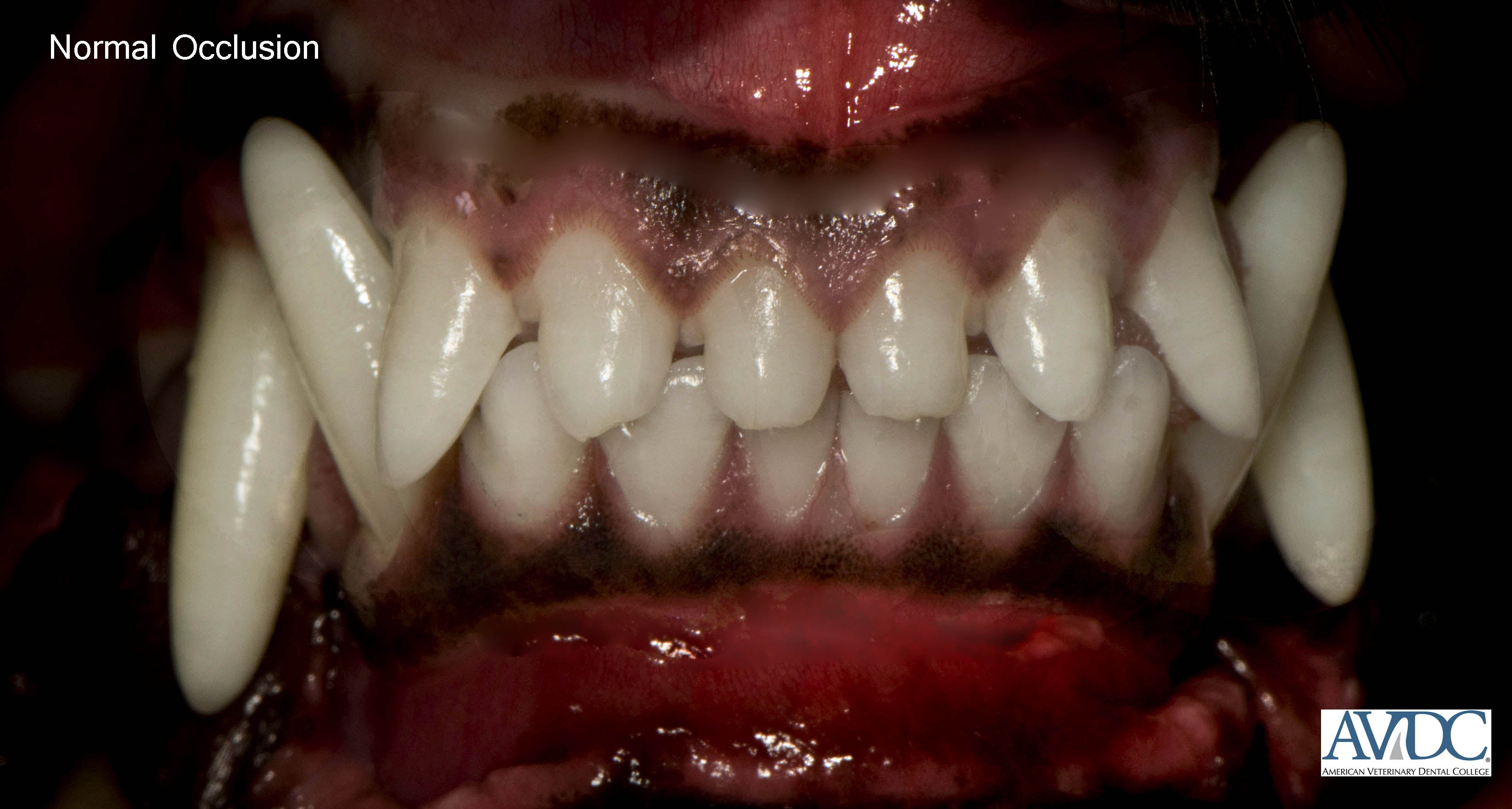
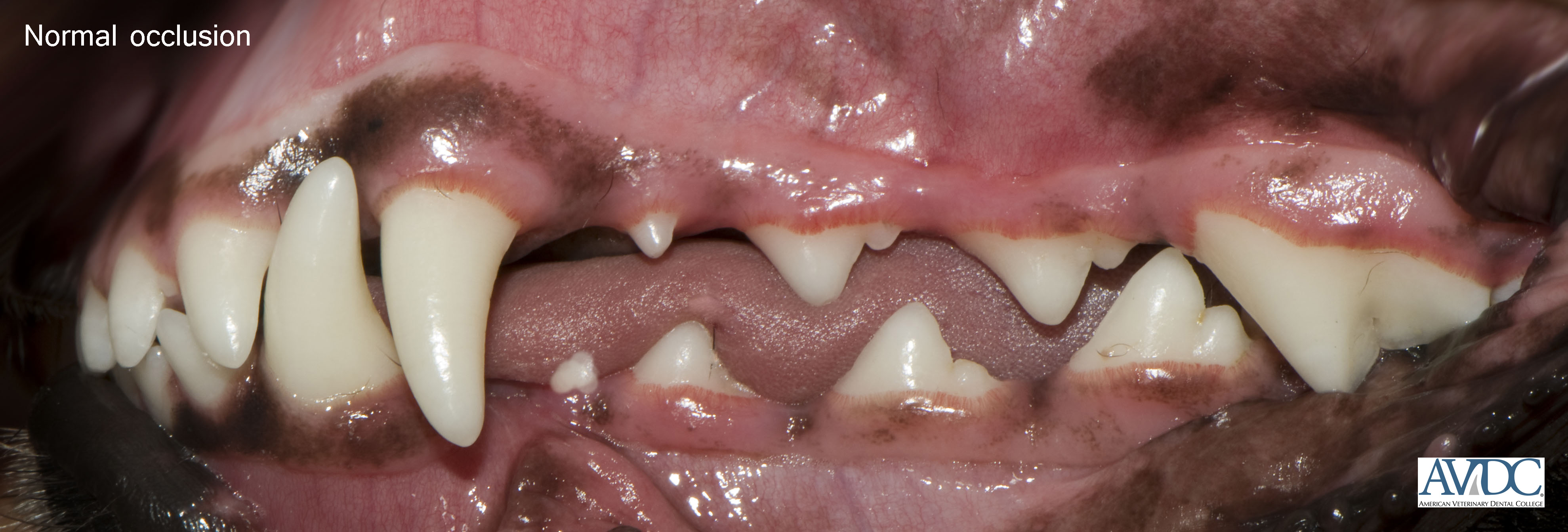
Normal Occlusion in a Cat:
Normal occlusion in cats is similar to dogs.
Maxillary incisor teeth are labial to the mandibular incisor teeth, with the incisal tips of the mandibular incisors contacting the cingula of the maxillary incisors or occluding just palatal to the maxillary incisors.
Mandibular canine teeth fit equidistant in the diastema between the maxillary third incisor teeth and the maxillary canine teeth, touching neither.
The incisor bite and canine interdigitation form the dental interlock.
Each mandibular premolar tooth is positioned mesial to the corresponding maxillary premolar tooth.
The maxillary second premolar tooth points in a space between the mandibular canine tooth and third premolar tooth.
The subsequent teeth interdigitate, with the mandibular premolars and first molar being situated lingual to the maxillary teeth.
The buccal surface of the mandibular first molar tooth occludes with the palatal surface of the maxillary fourth premolar tooth.
The maxillary first molar tooth is located distopalatal to the maxillary fourth premolar tooth.
Malocclusion
_________________________________________
Malocclusion (MAL) is any deviation from normal occlusion described above.
Malocclusion may be due to abnormal positioning of a tooth or teeth (dental malocclusion) or due to asymmetry or other deviation of bones that support the dentition (skeletal malocclusion).
The diagnosis for a patient with malocclusion is abbreviated as: MAL (malocclusion) 1 or 2 or 3 or 4 (= malocclusion class designation)/specific malocclusion abbreviation and tooth or teeth number(s).
Example 1: MAL1/CB/R202 for a dog with class 1 malocclusion and a rostral crossbite of the left maxillary second incisor.
If multiple teeth have the same malocclusion, include the tooth numbers with a comma in between e.g. MAL1/CB/R202,302
Example 2: MAL2/LV304 for a dog with class 2 malocclusion and linguoversion of the left mandibular canine tooth.
Dental Malocclusions
_________________________________________
Neutroclusion – Class 1 Malocclusion (MAL1):
A normal rostrocaudal relationship of the upper and lower dental arches with malposition of one or more individual teeth.
Distoversion(MAL1/DV) describes a tooth that is in its anatomically correct position in the dental arch but which is abnormally angled in a distal direction.
Mesioversion (MAL1/MV) describes a tooth that is in its anatomically correct position in the dental arch but which is abnormally angled in a mesial direction.
Linguoversion (MAL1/LV) describes a tooth that is in its anatomically correct position in the dental arch but which is abnormally angled in a lingual direction.
Palatoversion (MAL1/PV) describes a tooth that is in its anatomically correct position in the dental arch but which is abnormally angled in a palatal direction.
Labioversion (MAL1/LABV) describes an incisor or canine tooth that is in its anatomically correct position in the dental arch but which is abnormally angled in a labial direction.
Buccoversion (MAL1/BV) describes a premolar or molar tooth that is in its anatomically correct position in the dental arch but which is abnormally angled in a buccal direction.
Crossbite (CB) describes a malocclusion in which a mandibular tooth or teeth have a more buccal or labial position than the antagonist maxillary tooth. It can be classified as rostral or caudal:
In rostral crossbite (CB/R): One or more of the mandibular incisor teeth is labial to the opposing maxillary incisor teeth when the mouth is closed. Similar to posterior crossbite in human terminology.
In caudal crossbite (CB/C): One or more of the mandibular cheek teeth is buccal to the opposing maxillary cheek teeth when the mouth is closed. Similar to posterior crossbite in human terminology.
Skeletal Malocclusion
An abnormal relationship of the upper and lower dental arches; in addition to that there could also be malposition (i.e., -version) of one or more individual teeth.
Symmetrical Skeletal Malocclusions:
_________________________________________
Mandibular Distoclusion – Class 2 Malocclusion (MAL2):
An abnormal rostrocaudal relationship between the dental arches in which the lower dental arch occludes caudal to its normal position relative to the upper dental arch. Example:
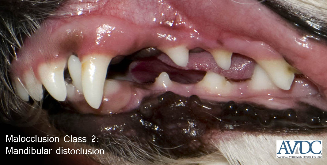
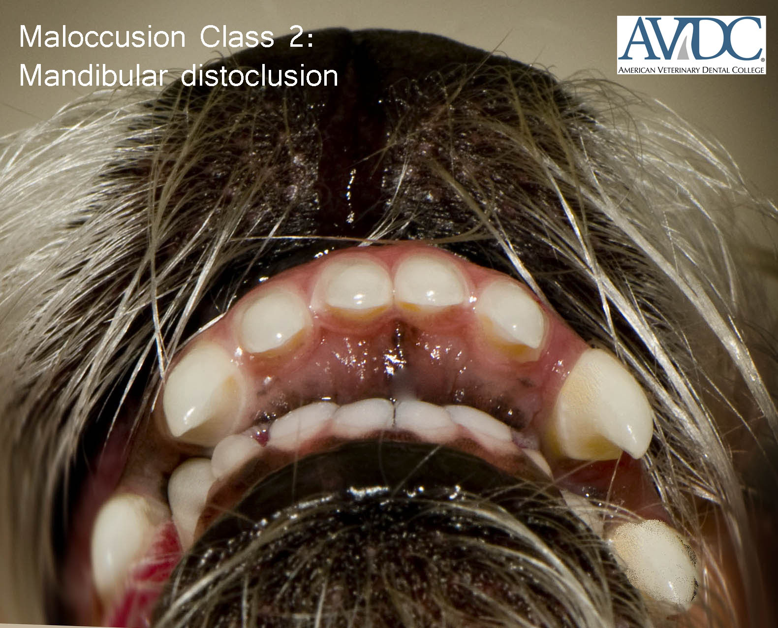
Mandibular mesioclusion – Class 3 Malocclusion: (MAL3)
An abnormal rostralcaudal relationship between the dental arches in which the lower dental arch occludes rostral to its normal position relative to the upper dental arch.
Example:
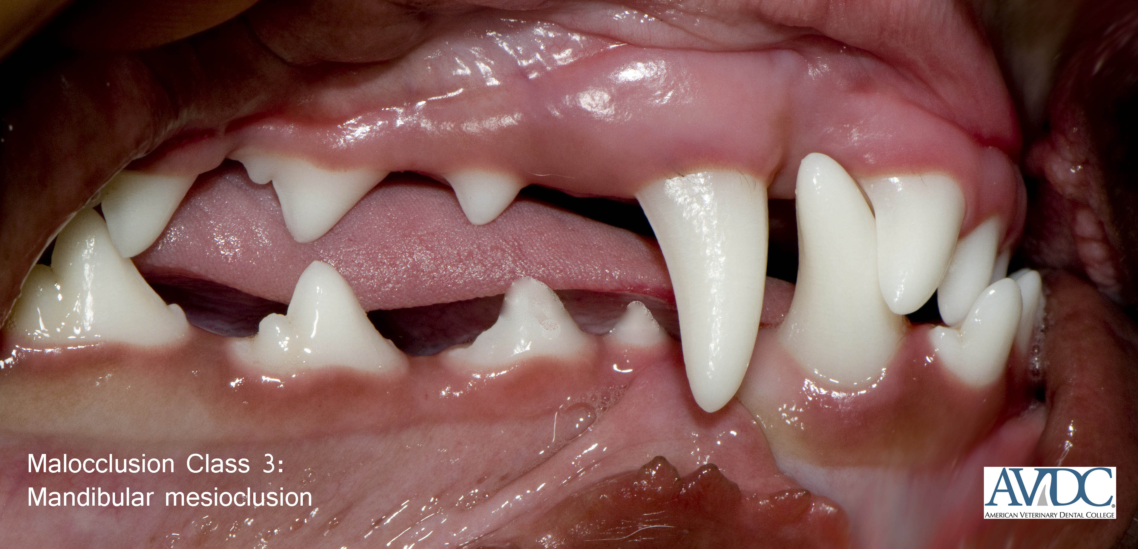
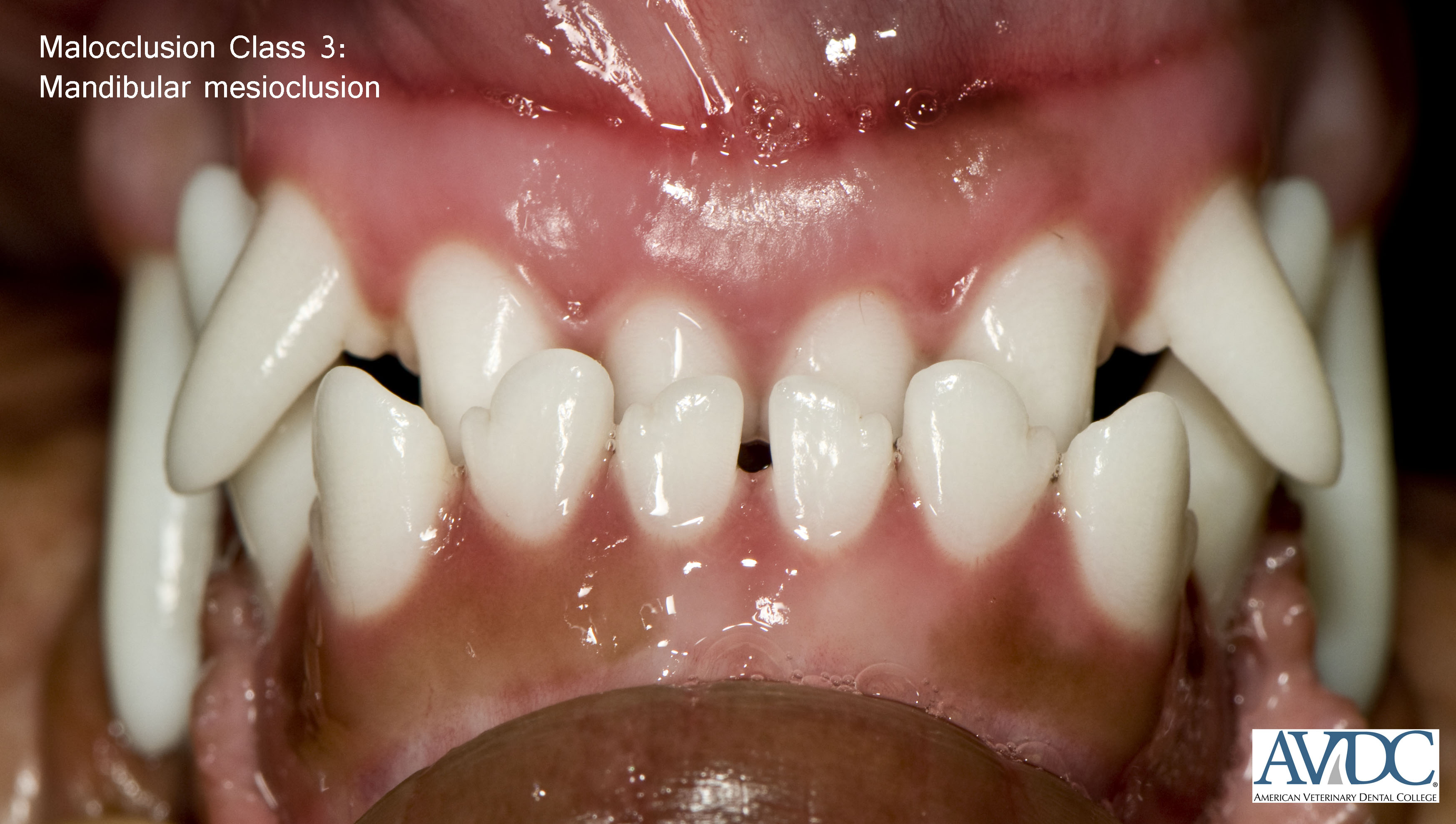
Asymmetrical Skeletal Malocclusions:
_________________________________________
Maxillomandibular Asymmetry – Class 4 Malocclusion: (MAL4)
A skeletal malocclusion with asymmetry in a rostrocaudal, side-to-side, or dorsoventral direction:
Maxillomandibular asymmetry in a rostrocaudal direction (MAL4/RC) occurs when mandibular mesioclusion or distoclusion is present on one side of the face while the contralateral side retains normal dental alignment.
Maxillomandibular asymmetry in a side-to-side direction (MAL4/STS) occurs when there is loss of the midline alignment of the maxilla and mandible.
Maxillomandibular asymmetry in a dorsoventral direction (MAL4/DV) results in an open bite, which is defined as an abnormal vertical space between opposing dental arches when the mouth is closed.
The expression “wry bite” is a layman term that has been used to describe a wide variety of unilateral occlusal abnormalities. Because “wry bite” is non-specific, its use is not recommended.
Management of Malocclusion
_________________________________________
Orthodontics
is a specialty in dentistry and oral surgery that is concerned with the prevention, interception and correction of malocclusion.
Preventive orthodontics
is concerned with the client’s education, the development of the dentition and maxillofacial structures, the diagnostic procedures undertaken to predict malocclusion and the therapeutic procedures instituted to prevent the onset of malocclusion. Preventive procedures are undertaken in anticipation of development of a problem. Examples of preventive procedures include:
• Client education about time tables on exfoliation of deciduous teeth and eruption of permanent teeth
• Fiberotomy (severing of gingival fibers around a permanent tooth to prevent its relapse after corrective orthodontics)
• Operculectomy (surgical removal of an operculum to enable eruption of a permanent tooth)
• Extraction of a tooth that could pose a risk to development of malocclusion
Interceptive orthodontics
is concerned with the elimination of a developing or established malocclusion. Interceptive procedures are typically undertaken in the growing patient. Examples of interceptive procedures include:
• Crown reduction of a permanent tooth in malocclusion
• Extraction of a tooth in malocclusion
Corrective orthodontics
is concerned with the correction of malocclusion without loss of the maloccluded tooth or part of its crown. This is accomplished by means of tooth movement. Examples of corrective procedures include:
• Surgical repositioning of a tooth
• Orthognathic surgery to treat skeletal malocclusion
• Passive movement of a tooth using an inclined plane
• Active movement of a tooth using an elastic chain
Treatment plan (TP):
Written document that outlines the progression of therapy (advantages, disadvantages, costs, alternatives, outcome and duration of treatment)
Impression (IM):
Detailed imprint of hard and/or soft tissues that is formed with specific types of impression materials
Full-mouth impression (IM/F):
Imprints of the dentition and/or surrounding soft tissues of the upper and lower dental arches
Diagnostic cast (DC):
Positive replica created by pouring a liquid material into an impression or placing an impression into a liquid material; once the material has hardened, the cast is removed and used for the purpose of study and treatment planning; also called die
(DC/D)
when made from an impression of a particular tooth/area of interest or stone model
(DC/SM)
when made from a full-mouth impression
Bite registration (BR):
Impression used to record a patient’s occlusion, which is then used to articulate diagnostic casts
Fiberotomy (FT):
Severing gingival fibers around a permanent tooth to prevent its relapse after corrective orthodontics
Operculectomy (OP):
Surgical removal of an operculum to enable eruption of a permanent tooth
Surgical repositioning (SR):
Repositioning of a developmentally displaced tooth
Orthognathic surgery (OS):
Surgical procedure to alter relationships of dental arches typically performed to correct skeletal malocclusion
Bracket/button/hook (OA/BKT):
Device made of metal or plastic that is bonded to the tooth surface and aids in the attachment of wires or elastics; use
OA/CMB
if custom-made
Elastic chain/tube/thread (OA/EC):
Orthodontic elastics used to move teeth
Orthodontic wire (OA/WIR):
Metal wire with ‘memory’ used to move teeth
Arch bar (OA/AR):
Device attached to one dental arch to move individual teeth in between the device’s attachments
Orthodontic appliance (OA):
Device attached to a tooth or teeth to move a tooth or teeth
Orthodontic appliance adjustment (OA/A):
Abbreviation used at the time of adjustment of the orthodontic appliance
Orthodontic appliance installation (OA/I):
Abbreviation used at the time of installation of the orthodontic appliance
Orthodontic appliance removal (OA/R):
Abbreviation used at the time of removal of the orthodontic appliance
Orthodontic counseling (OC):
Client communication on the genetic basis, diagnosis and treatment of malocclusion and the legal and ethical implications of orthodontics
Ball therapy (BTH):
Removable orthodontic device in the form of a ball or cone-shaped rubber toy (for example to passively move linguoverted mandibular canine teeth)
Inclined plane (IP):
Fixed orthodontic device made of acrylic (IP/A), composite (IP/C) or metal (IP/M) with sloping planes (for example to passively move linguoverted mandibular canine teeth)
Orthodontic recheck (OR): Examination of a patient treated with an orthodontic appliance.
Tooth Extraction-Related Terminology
Palate, Pharyngeal and Nasal Surgery
Grafts and Related Terminology
——–
Tooth Extraction-Related Terminology
_________________________________________
Closed extraction (X or XS):
Extraction of teeth without flap creation; X is used when closed extraction is performed without tooth sectioning; XS is used when closed extraction is performed with tooth sectioning or removal of interproximal crown tissue
Open extraction (XSS):
Extraction of teeth after flap creation and alveolectomy
Alveolectomy (ALV):
Removal of some or all of the alveolar bone
Alveoloplasty (ALV):
A form of alveolectomy performed to restore physiological contours or achieve smooth contours of the alveolar bone.
Palate, Pharynx and Nasal Surgery
_________________________________________
Palate Surgery – Click the link
Naroplasty (NAS/R):
Surgical correction of stenotic nares
Tonsillectomy (TON/X):
Surgical resection of the palatine tonsil
Grafts and Related Terminology
_________________________________________
Transplantation:
Act or process of transferring something from one part or individual to another.
Transplant:
Something that is transferred from one part or individual to another.
Graft (GF):
Non-living material or living tissue used for implantation or transplantation to replace a diseased part or compensate for a defect.
Gingival graft (GF/G):
Gingiva or gingiva-like tissue (e.g., from the hard palate) used to replace gingiva in a gingival defect.
Connective tissue graft (GF/CT):
Connective tissue from a keratinized mucosa (e.g., from the hard palate) placed in a gingival defect and which is partially or completely covered with gingiva and/or alveolar mucosa in the recipient bed.
Mucosal graft (GF/M)
Mucosa used to take place of a removed piece of mucosa or cover a mucosal defect.
Bone graft (GF/B):
A surgical procedure by which bone or a bone substitute is used to take place of a removed piece of bone or bony defect.
Cartilage graft (GF/C):
Cartilage used to to take the place of a removed piece of bone or fill a bony defect.
Skin graft (GF/S):
Skin used to take place of a removed piece of skin/mucosa or skin/mucosa defect.
Venous graft (GF/V):
A vein used to take place of a removed segment of artery/vein or arterial/venous defect.
Nerve graft (GF/N):
A nerve used to take place of a removed segment of nerve or nerve defect.
Fat graft (GF/F):
Adipose tissue used to provide volume to a defect or to prevent ingrowth of other tissues into the defect.
Autograft:
Tissue transferred from one area to another area of the animal’s own body.
Isograft:
Tissue transferred between genetically identical animals.
Allograft:
Tissue transferred between genetically dissimilar animals of the same species.
Xenograft:
Tissue transferred between animals of different species.
Particulate graft:
A graft containing equally or variably-sized particles.
Full-thickness graft:
A graft consisting of the full thickness of a tissue.
Partial-thickness (split-thickness) graft:
A graft consisting of a portion of the thickness of a tissue.
Mesh graft:
A type of partial-thickness graft in which multiple small incisions have been made to increase stretching and flexibility of the graft.
Composite graft:
A graft composed of at least two different tissues, e.g., skin-muscle-and-bone graft.
Implant (IMP):
Something inserted into or applied onto living tissue.
Implantation:
The act or process of inserting something into or applying something onto living tissue.
Equine Anatomical Structures
Equine Dental Abnormalities
Equine Tooth Extractions
——–
Equine Dental Anatomical Structures
_________________________________________
Infundibulum (INF): Enamel cup-like infolding from the occlusal surface in incisors (one) and maxillary cheek teeth (two)
Diastema (D): Space between teeth in a jaw.
Pulp horn (PH): An elongation of the pulp chamber extending towards the cusps of brachyodont teeth; an elongation of the common pulp chamber extending towards the occlusal surface of equine cheek teeth (numbers refer to the Du Toit numbering system, e.g., PH1, PH2, etc.).
Regular secondary dentin: Dentin produced on the periphery of the pulp after the tooth has come into occlusion which gradually reduces the size of the pulp horns
Irregular secondary dentin: Physiological dentin that is laid down last, sub-occlusally in the center of the pulp horn which prevents pulp exposure with normal tooth wear
Sinus (SIN): Paranasal cavity within a bone
Conchofrontal sinus (SIN/CF): Compound term for the frontal sinus and the dorsal conchal sinus, which are continuous in equines
Caudal maxillary sinus (SIN/CMX): Cavity in equines separated by the maxillary septum from the rostral maxillary sinus; communicating with the frontal and sphenopalatine sinuses
Rostral maxillary sinus (SIN/RMX): Cavity in equines separated by the maxillary septum from the caudal maxillary sinus; opening freely into the ventral conchal sinus
Sphenopalatine sinus (SIN/SP): Continued cavity formed by the sphenoid and palatine sinuses in equines; opening into the caudal maxillary sinus
Ventral conchal sinus (SIN/VC): Cavity enclosed by the caudal part of the ventral concha.
Equine Dental Abnormalities and Procedures
_________________________________________
Tooth elongation (T/EL): Abnormal intraoral and/or periapical extension of the coronal and/or apical portion of the tooth
Open diastema (D/O): Pathological widening of the interproximal space that has similar widths at the gingival margin and occlusal surface
Valve diastema (D/V): Pathological widening of the interproximal space that is considerably wider at the gingival margin than at the occlusal surface
Temporal teratoma (TT): Vestigial dental structure in the vicinity of the temporal bone as a result of failure of closure of the first branchial cleft usually associated with swelling or a sinus tract at the base of the pinna of the ear; also known as heterotopic polyodontia or “ear tooth” and erroneously called dentigerous cyst in the horse
Pulp horn defect (PH/D): Pulp horn exposure or defective secondary dentin overlying a pulp horn noted on the occlusal surface of cheek teeth which may or may not be vital
Infundibular caries (CA/INF): Caries of the maxillary cheek teeth infundibulae; grade 1 involving cementum only; grade 2 also involving enamel; grade 3 also involving dentin; grade 4 affecting the structural integrity of the tooth; use CA/INF/D for distal infundibular caries and CA/INF/M for mesial infundibular caries
Peripheral caries (CA/PER): – Caries affecting the periphery of the cheek teeth; grade 1 involving cementum only; grade 2 also involving enamel; grade 3 also involving dentin; grade 4 affecting the structural integrity of the tooth
Shear mouth (SHE): Abnormally increased occlusal angulation of the cheek teeth; for example >40° for mandibular cheek teeth and >25° for maxillary cheek teeth
Sinusitis (SIN/IN): Inflammation of the sinus (e.g. SIN/IN/RMX = rostral maxillary sinusitis)
Primary sinusitis: Inflammation of the sinus associated with bacterial infection of the sinuses without any detectable predisposing cause.
Secondary sinusitis: Inflammation of the sinus associated with bacterial infection where a predisposing cause such as periapical disease or intra-sinus growth is present
Dental sinusitis: Sinusitis caused by periapical disease of the caudal cheek teeth
Diastema odontoplasty (or widening) (D/ODY): Removal of interproximal dental tissue to avoid entrapment of food between teeth
Sinoscopy (SIN/EN): Endoscopic examination of the sinus using a trephine portal
Sinus flap (SIN/F): Surgical access to the sinus via a skin and bone flap; use SIN/CF/F for a chonchofrontal sinus flap and SIN/MX/F for a maxillary sinus flap
Sinus lavage (SIN/LAV): Flushing of the sinus
Equine Tooth Extraction and Related Procedures
_________________________________________
Trephination (TRP): Surgical access to a structure of interest via skin incision and removal of a circular piece of bone using a trephine
Sinus trephination (SIN/TRP): Access to the sinus via a trephined hole
Closed extraction with odontoplasty (XS/ODY): Removal of interproximal crown tissue to facilitate transoral extraction of a tooth
Extraction of a tooth after apicoectomy and repulsion (XSS/APX/RPL): Extraction of a tooth after apicoectomy and repulsion which is performed via TRP, SIN/TRP or SIN/F
Transbuccal extraction (XSS/BUC): Extraction of a tooth after buccotomy
Transbuccal extraction with alveolectomy (XSS/BUC/ALV): Extraction of a tooth after buccotomy and alveolectomy
Transcommissural extraction (XSS/COM): Extraction of a tooth after commissurotomy
Transcommissural extraction with alveolectomy (XSS/COM/ALV): Extraction of a tooth after commissurotomy and alveolectomy
Minimally invasive buccotomy extraction (XSS/MIB): Extraction of a tooth via minimally invasive buccotomy (small incision made for introduction of straight instrumentation to elevate, section or drill into a cheek tooth for the purpose of facilitating its transoral extraction)
Extraction via repulsion (XSS/RPL): Extraction of a tooth after repulsion which is performed via TRP, SIN/TRP or SIN/F
English is the official language of AVDC®. All documents, forms and examinations provided by AVDC® are written in English, and all materials submitted by applicants and residents are to be written in English.
Use of Specialty Titles: The AVMA Principles of Veterinary Medical Ethics state: It is unethical for veterinarians to identify themselves as or in any way imply that they are members of an AVMA recognized specialty organization until such time as such certification has been awarded. AVDC® has adopted the Guidelines for Use of Specialty Titles, prepared by the American Board of Veterinary Specialties of the American Veterinary Medical Association.
By signing the AVDC® Training Program Registration form, an individual submitting an application for registration of a training program acknowledges that s/he may not use the terms ‘board eligible’, ‘board qualified’, ‘veterinary dental specialist’ or ‘veterinary dentist’, and may not make or imply any connection with AVDC® until such time as the individual passes the AVDC® examination and is awarded the AVDC® Diploma.
The registration of an AVDC® training program of an individual who is not in compliance with the policy stated above may be terminated by AVDC®, in which case the individual will not be permitted to complete the AVDC® credentials and/or the examination procedures, and, when appropriate, the circumstances may be reported by AVDC® to the individual’s State Board of Veterinary Medical Examiners.